Copyright
©The Author(s) 2017.
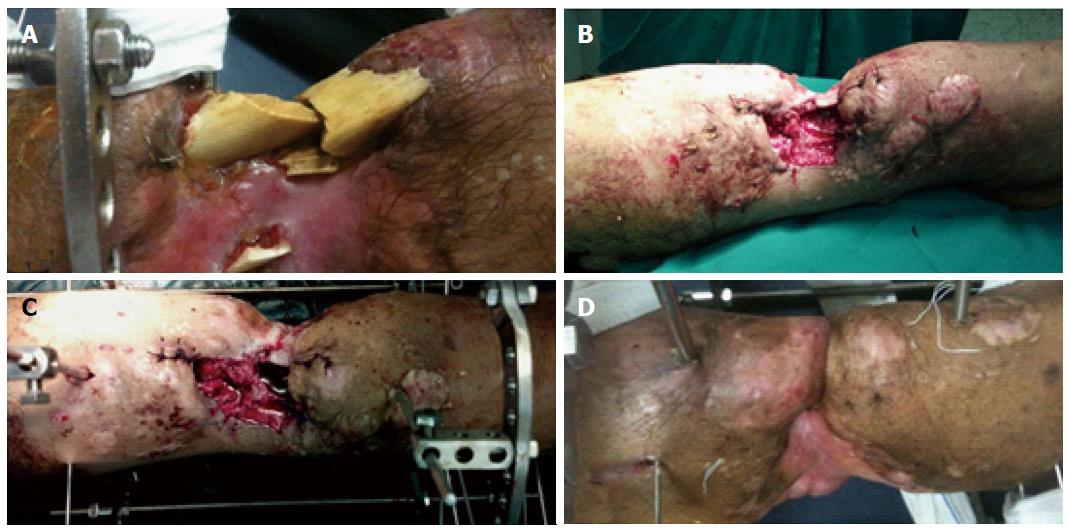
Figure 1 Surgical technique.
The infected and necrotic bones are excised (A) and further debridement of the bone ends is done until they recessed under the skin and soft tissues (B); C: The Ilizarov frame is applied and bone transport started; D: At the time of docking the skin is invaginated between the bone ends.
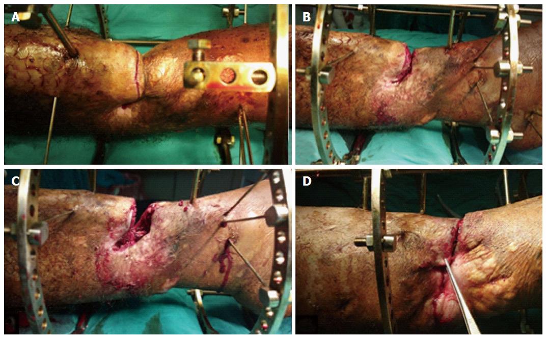
Figure 2 Management of the docking site.
A: Transverse skin incision is made along the bone ends at the docking site; B: The incision is deepened down to the bone and the soft tissues are removed from the docking site; C: The bone ends are freshened and compressed against each other by the frame; D: This compression approximates the skin edges together and facilitates their closure over the bone ends.
Figure 3 Middle third bone and soft tissue defects.
A: Forty-seven year old male patient with combined bone loss, soft tissue loss and infection of his left leg as a complication of an open fracture of the tibia and fibula; B: Debridement of the bone ends was done, Ilizarov frame applied and bone transport started; C: During bone transport the soft tissue defect gradually closes; D: At the time of docking the skin was fashioned over the bone ends; E: After removal of the frame with good bone healing; F: The patient with good alignment and complete healing of the soft tissue.
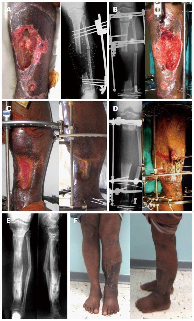
Figure 4 Lower third bone and soft tissue defects.
A: Thirty-nine year old male patient with skin loss, infection and exposed plate in the lower third of his right leg; B: After radical debridement and application of the Ilizarov external fixator; At the time of docking the skin was fashioned to cover the bone ends (C) and it healed completely (D); After removal of the frame with good bone healing (E) and the patient with good alignment and complete healing of the soft tissue (F).
- Citation: El-Alfy BS. Unhappy triad in limb reconstruction: Management by Ilizarov method. World J Orthop 2017; 8(1): 42-48
- URL: https://www.wjgnet.com/2218-5836/full/v8/i1/42.htm
- DOI: https://dx.doi.org/10.5312/wjo.v8.i1.42


