Copyright
©The Author(s) 2016.
World J Orthop. Jul 18, 2016; 7(7): 458-462
Published online Jul 18, 2016. doi: 10.5312/wjo.v7.i7.458
Published online Jul 18, 2016. doi: 10.5312/wjo.v7.i7.458
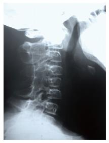
Figure 1 Plain X-rays at the age of 12 of cervical spine showed loss of lordosis and posterior fusion from C2 to C6 joints.
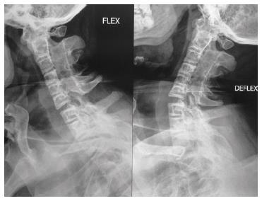
Figure 2 Bending films on admission showed spontaneous apophyseal joint fusion from the occipital condyle to C6 and from C7 to Th2 with marked instability between C6 and C7.
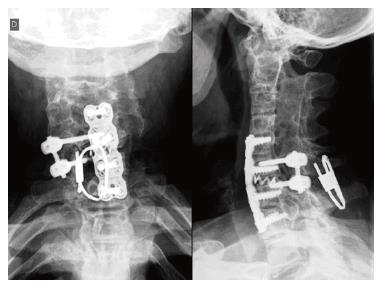
Figure 3 Post-operative plain X-rays.
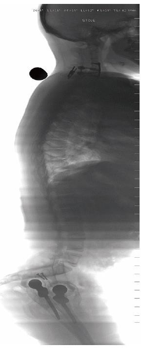
Figure 4 Post-operative scan; showing restored sagittal alignment.
- Citation: Suhodolčan L, Mihelak M, Brecelj J, Vengust R. Operative stabilization of the remaining mobile segment in ankylosed cervical spine in systemic onset - juvenile idiopathic arthritis: A case report. World J Orthop 2016; 7(7): 458-462
- URL: https://www.wjgnet.com/2218-5836/full/v7/i7/458.htm
- DOI: https://dx.doi.org/10.5312/wjo.v7.i7.458


