Copyright
©The Author(s) 2016.
World J Orthop. Mar 18, 2016; 7(3): 167-170
Published online Mar 18, 2016. doi: 10.5312/wjo.v7.i3.167
Published online Mar 18, 2016. doi: 10.5312/wjo.v7.i3.167
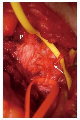
Figure 1 Photo taken during the surgical release of a left-side middle cluneal nerve.
Medially to the posterior superior iliac crest (P), the MCN is identified passing under the superficial layer of the long posterior sacroiliac ligament. The nerve is seen to be entrapped under the deeper layer of the long posterior sacroiliac ligament where it penetrates the ligament (arrow). The yellow and red tapes have been used to lift the proximal and distal portions of MCN branch, respectively. MCN: Middle cluneal nerve.
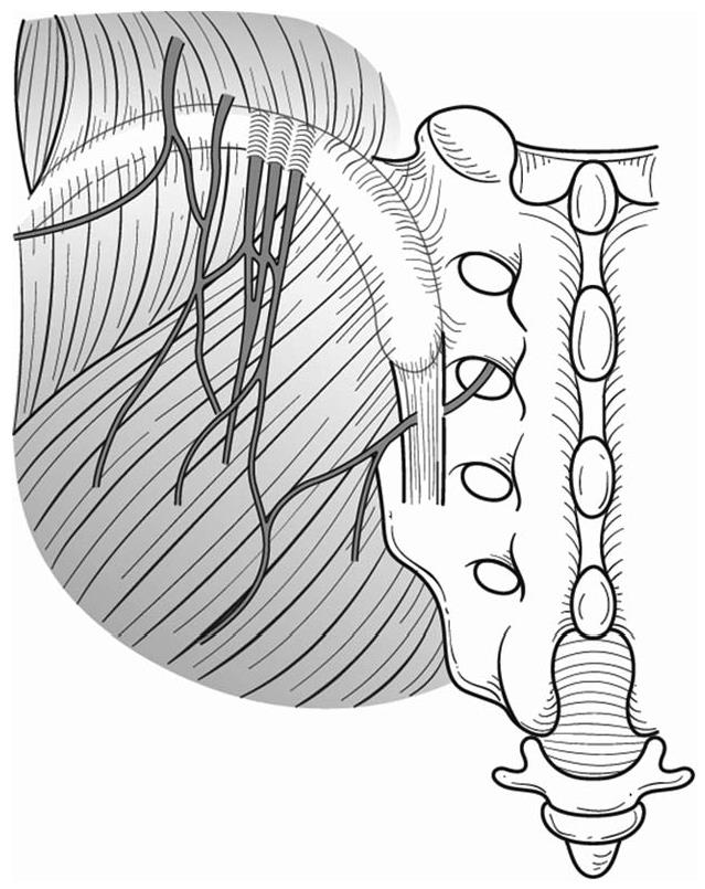
Figure 2 Schematic illustration of typical running courses and entrapment of superior and middle cluneal nerves.
Multiple branches of the superior cluneal nerve may be entrapped where they pierce the thoracolumbar fascia over the iliac crest. Middle cluneal nerve may be entrapped where this nerve pass under or through the long posterior sacroiliac ligament.
- Citation: Aota Y. Entrapment of middle cluneal nerves as an unknown cause of low back pain. World J Orthop 2016; 7(3): 167-170
- URL: https://www.wjgnet.com/2218-5836/full/v7/i3/167.htm
- DOI: https://dx.doi.org/10.5312/wjo.v7.i3.167


