Copyright
©The Author(s) 2015.
World J Orthop. Nov 18, 2015; 6(10): 847-849
Published online Nov 18, 2015. doi: 10.5312/wjo.v6.i10.847
Published online Nov 18, 2015. doi: 10.5312/wjo.v6.i10.847
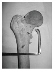
Figure 1 Langenbeck retractor is inserted with its blade tip facing proximally and advanced well beyond the medial cortex of femur (in this example a 15 mm × 45 mm retractor is shown, in clinical practice a 10 mm x 30 mm or smaller retractor is preferable).
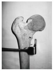
Figure 2 Retractor blade is rotated by 90°.
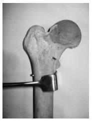
Figure 3 Drill bit is pushed back by the blade of the retractor.
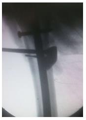
Figure 4 Image intensifier view of the technique.
- Citation: Chouhan DK, Sharma S. “Push back” technique: A simple method to remove broken drill bit from the proximal femur. World J Orthop 2015; 6(10): 847-849
- URL: https://www.wjgnet.com/2218-5836/full/v6/i10/847.htm
- DOI: https://dx.doi.org/10.5312/wjo.v6.i10.847


