Copyright
©The Author(s) 2024.
World J Orthop. May 18, 2024; 15(5): 477-482
Published online May 18, 2024. doi: 10.5312/wjo.v15.i5.477
Published online May 18, 2024. doi: 10.5312/wjo.v15.i5.477
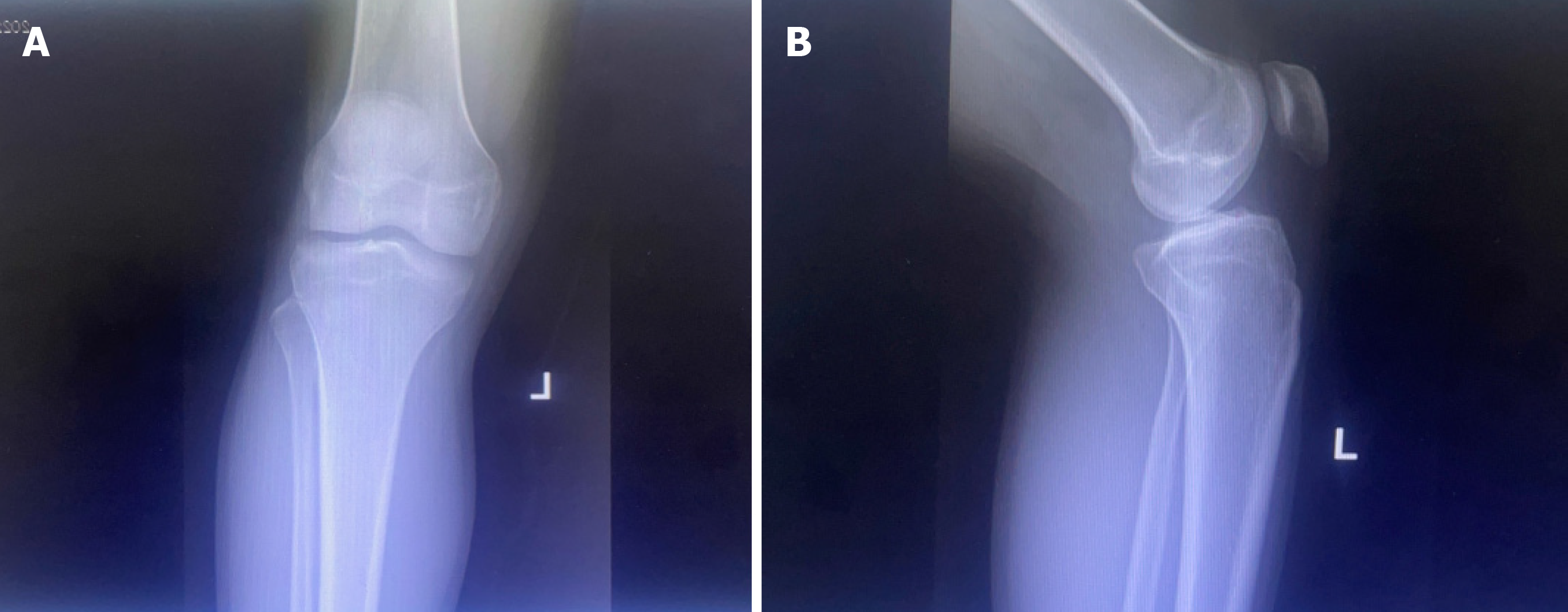
Figure 1 Plain radiography of the left knee displays preserved joint spaces with no fractures or dislocations.
A: Plain anteroposterior radiography; B: Plain lateral radiography.
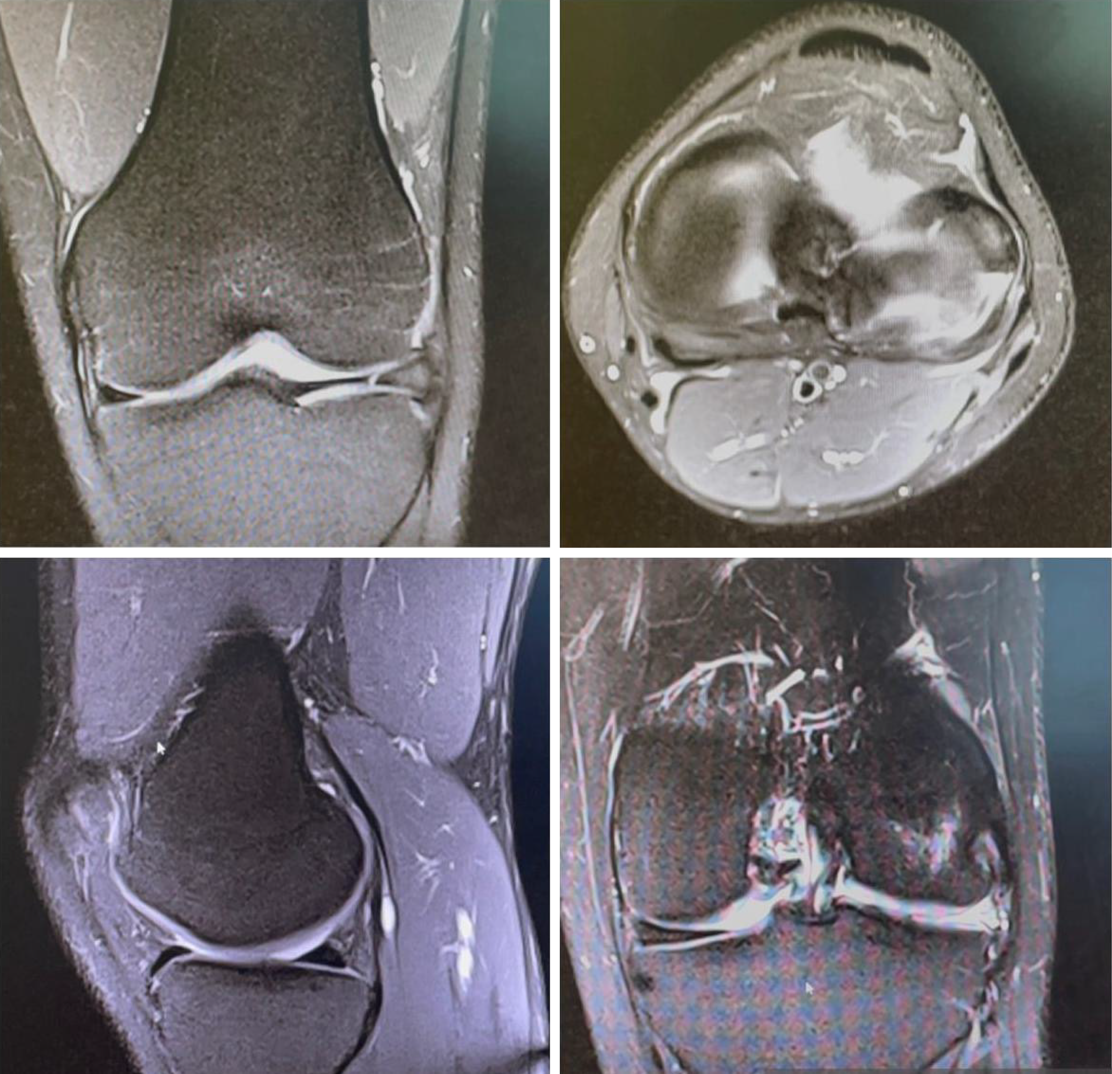
Figure 2
Coronal, axial, and sagittal T2-weighted magnetic resonance imaging of the left knee demonstrates intact cruciate ligaments and medial meniscus.
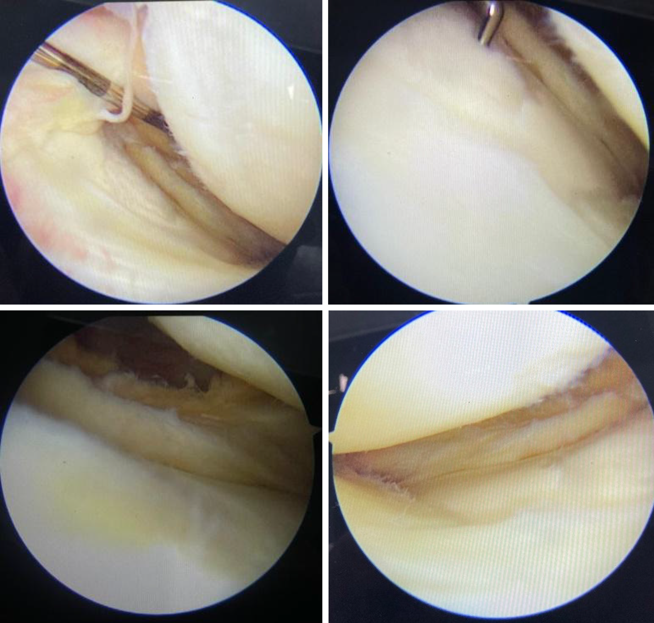
Figure 3
Arthroscopic views of the left knee display a congenital absence of the lateral meniscus.
- Citation: Alkhunayfir HA, AlQahtani AA, Korkoman AJ. Congenital absence of the lateral meniscus: A case report. World J Orthop 2024; 15(5): 477-482
- URL: https://www.wjgnet.com/2218-5836/full/v15/i5/477.htm
- DOI: https://dx.doi.org/10.5312/wjo.v15.i5.477


