Copyright
©The Author(s) 2023.
World J Orthop. Jul 18, 2023; 14(7): 562-571
Published online Jul 18, 2023. doi: 10.5312/wjo.v14.i7.562
Published online Jul 18, 2023. doi: 10.5312/wjo.v14.i7.562
Figure 1 Intraoperative fluroscopy images used to insert the internal fixator screws.
A: Obturator outlet view showing the teardrop (*). A sharp starting awl was used to open the cortex for screw insertion; B: Pedicle finder was then used to establish the bony tunnel. Correct screw trajectory was checked in the obturator outlet, obturator inlet and iliac views; C: After screw insertion the three views checked agin to ensure that the screw did not penetrate the cortex and was placed totally intraosseous.
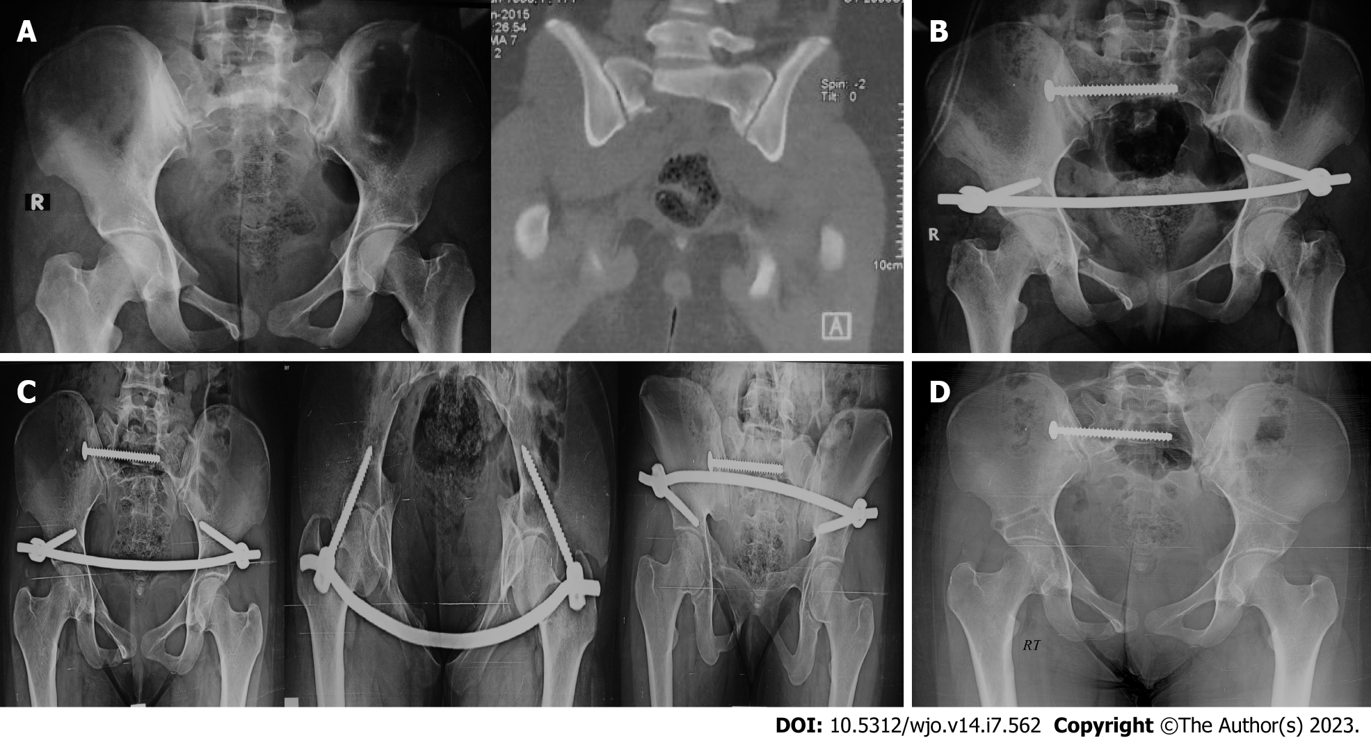
Figure 2 17-year-old girl sustained a Tile C type pelvic fracture after fall from a height of 6 m.
A: Pelvis antero-posterior (AP) X-ray view and sagittal computed tomography scans showing sacral and ramus fractures on the Rt. Side; B: The pelvic fracture was fixed using a fully threaded sacroiliac screw and an internal fixator (INFIX); C: 3-mo follow-up pelvic X-ray (AP, inlet and outlet) views showing complete union. The INFIX was removed 2 mo later in the OR; D: One-year follow-up X-ray. The patient had excellent function with complete return to her prefracture activity level.
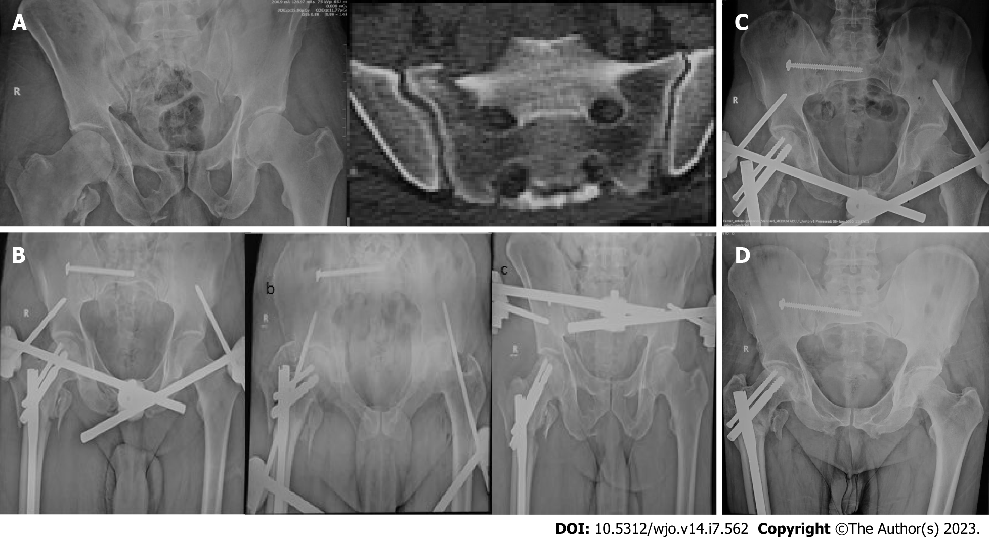
Figure 3 57-year-old male patient sustained a Tile B type pelvic fracture after fall from a 3 meter height on his Rt.
side. A: Pelvis antero-posterior (AP) X-ray view and axial computed tomography cuts showing Rt. sacral and bilateral rami fractures. The patient has an associated Rt. pertrochanteric fracture; B: The pelvic fracture was fixed using a fully threaded sacroiliac screw and an external fixator (EXFIX). The pertrochanteric fracture was also fixed at the same session using a long femoral nail; C: 3-mo follow-up pelvic X-ray (AP, inlet and outlet) views showing complete union. The EXFIX was removed in the outpatient clinic on the same day; D: 9-mo follow-up X-ray showing consolidation of the fractures. The patient had excellent function with complete return to his prefracture activity level.
- Citation: Abo-Elsoud M, Awad MI, Abdel Karim M, Khaled S, Abdelmoneim M. Internal fixator vs external fixator in the management of unstable pelvic ring injuries: A prospective comparative cohort study. World J Orthop 2023; 14(7): 562-571
- URL: https://www.wjgnet.com/2218-5836/full/v14/i7/562.htm
- DOI: https://dx.doi.org/10.5312/wjo.v14.i7.562


