Copyright
©The Author(s) 2022.
World J Orthop. Mar 18, 2022; 13(3): 259-266
Published online Mar 18, 2022. doi: 10.5312/wjo.v13.i3.259
Published online Mar 18, 2022. doi: 10.5312/wjo.v13.i3.259
Figure 1 Classification of the transverse relaxation (T2)-weighted images of magnetic resonance arthrography into three groups.
No leakage: Right shoulder injection into the glenohumeral joint without leakage; Minor leakage: Practical intra-articular right shoulder injection; however, another magnetic resonance arthrography image shows the presence of some leakage (white arrow) outside the posterior cuff; Major leakage: Inaccurate injection into the glenohumeral joint of the right shoulder with a noted severe/mass leakage (white arrow) surrounding the axillary area.
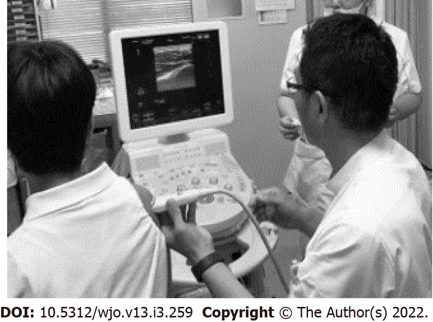
Figure 2 The setting of the injection procedure.
Glenohumeral injection performed by a shoulder surgeon using ultrasonic guidance. The patient sits upright with the shoulder at a neutral rotation position, and the ultrasonic probe is placed over the posterior part of the right shoulder.
Figure 3 Identification of the glenohumeral joint and needle insertion point on a captured ultrasound image.
A: The gap between the humeral head (H) and glenoid rim (G) is the target of this injection. The needle insertion point (asterisk) should be visualized more clearly; B: Ultrasonographic image during injection; C: Arrowhead shows the high echoic flow of the injection and arrow line indicates the assumed needle path.
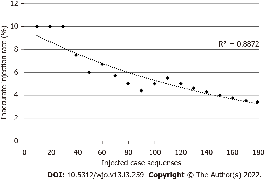
Figure 4 Learning curve.
The inaccurate injection rate is the total number of “major leakage” divided by the total number of injected cases that was recorded every 10 cases. The learning curve is represented by the dotted curve. Spearman’s rank correlation coefficient was analyzed (R2 = 0.887, P < 0.001).
- Citation: Kuratani K, Tanaka M, Hanai H, Hayashida K. Accuracy of shoulder joint injections with ultrasound guidance: Confirmed by magnetic resonance arthrography. World J Orthop 2022; 13(3): 259-266
- URL: https://www.wjgnet.com/2218-5836/full/v13/i3/259.htm
- DOI: https://dx.doi.org/10.5312/wjo.v13.i3.259


