Copyright
©The Author(s) 2022.
World J Orthop. Mar 18, 2022; 13(3): 250-258
Published online Mar 18, 2022. doi: 10.5312/wjo.v13.i3.250
Published online Mar 18, 2022. doi: 10.5312/wjo.v13.i3.250
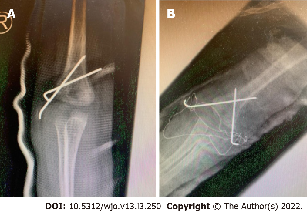
Figure 1 Postoperative AP view radiographs.
A: Lateral pinning; B: Cross pinning.
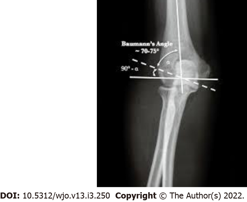
Figure 2 Lateral radiograph demonstrating Baumann's angle (angle between the long axis of humeral shaft and growth plate of lateral humeral condyle).
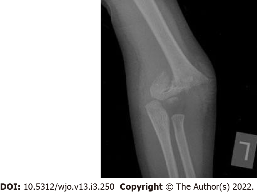
Figure 3 Pre-operative AP view radiograph.
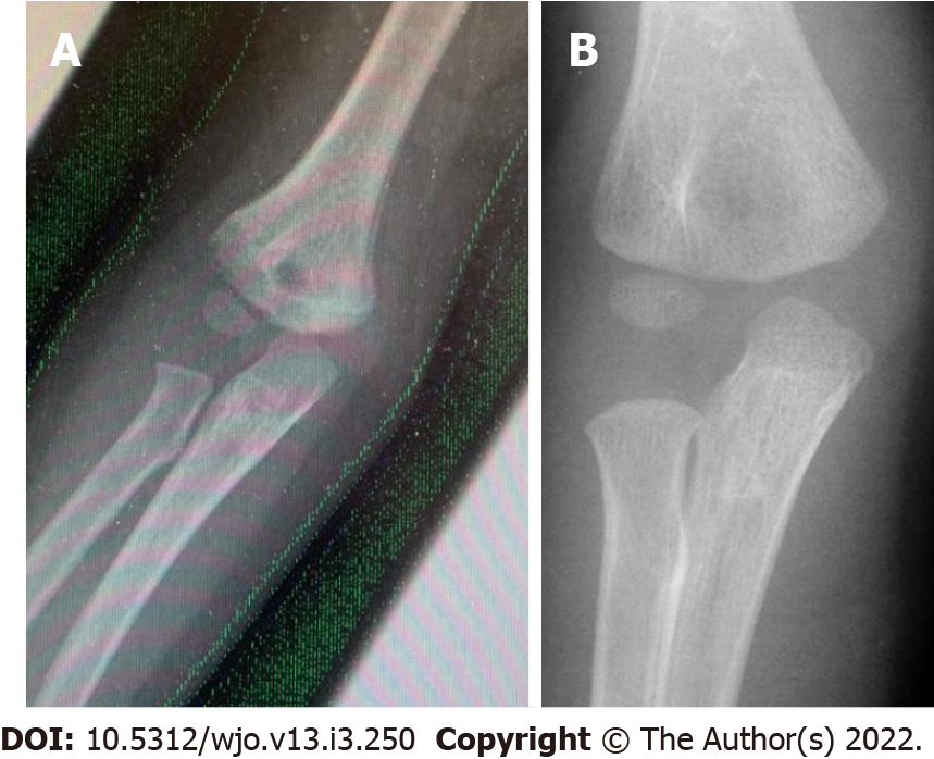
Figure 4 AP radiographs post pin removal.
A: Lateral pinning B: Crossed pinning.
- Citation: Radaideh AM, Rusan M, Obeidat O, Al-Nusair J, Albustami IS, Mohaidat ZM, Sunallah AW. Functional and radiological outcomes of different pin configuration for displaced pediatric supracondylar humeral fracture: A retrospective cohort study. World J Orthop 2022; 13(3): 250-258
- URL: https://www.wjgnet.com/2218-5836/full/v13/i3/250.htm
- DOI: https://dx.doi.org/10.5312/wjo.v13.i3.250


