Copyright
©The Author(s) 2021.
World J Orthop. Nov 18, 2021; 12(11): 945-953
Published online Nov 18, 2021. doi: 10.5312/wjo.v12.i11.945
Published online Nov 18, 2021. doi: 10.5312/wjo.v12.i11.945
Figure 1 Chest X-ray and computed tomography scan revealed right-sided pneumothorax.
A: Chest X ray in anterior-posterior view; B: Computed tomography scan in axial plane.
Figure 2 Computed tomography scan shows exostoses of five ribs.
The right first and seventh ribs were sharp and protruded into the thoracic cavity. A: Rt. (right) first rib; B: Rt. third rib; C: Rt. fourth rib; D: Rt. seventh; E: Left ninth rib. Rt.: Right.
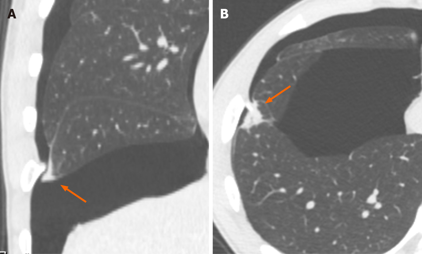
Figure 3 Damaged pleura and lung tissues confronted with the exostosis of the right seventh rib.
A: Coronal plane, thickness of visceral pleura; B: Axial plane, damaged lung tissues.
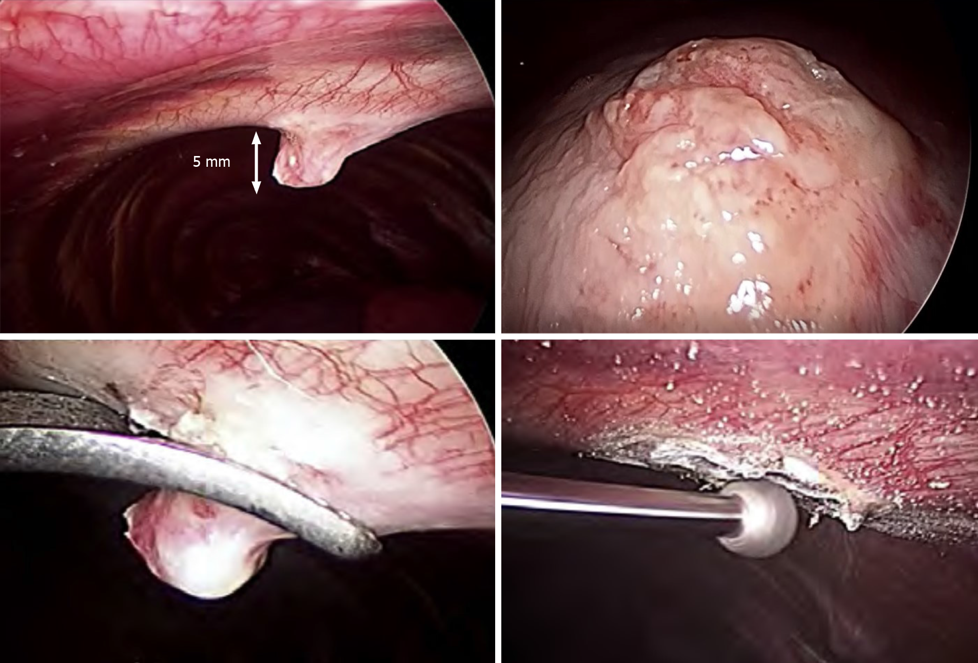
Figure 4 Intraoperative findings and treatment.
The bony spur was removed using forceps. The spinous lesion of the rib surface was scraped down by arthroscopic burr.
Figure 5 Pathological findings (hematoxylin and eosin stain, ×200).
The resected specimen was histologically composed of mature bone, hyaline cartilage, and fibrocartilage with no sign of malignant transformation.
- Citation: Nakamura K, Asanuma K, Shimamoto A, Kaneda S, Yoshida K, Matsuyama Y, Hagi T, Nakamura T, Takao M, Sudo A. Spontaneous pneumothorax in a 17-year-old male patient with multiple exostoses: A case report and review of the literature. World J Orthop 2021; 12(11): 945-953
- URL: https://www.wjgnet.com/2218-5836/full/v12/i11/945.htm
- DOI: https://dx.doi.org/10.5312/wjo.v12.i11.945


