Copyright
©The Author(s) 2021.
World J Orthop. Nov 18, 2021; 12(11): 867-876
Published online Nov 18, 2021. doi: 10.5312/wjo.v12.i11.867
Published online Nov 18, 2021. doi: 10.5312/wjo.v12.i11.867
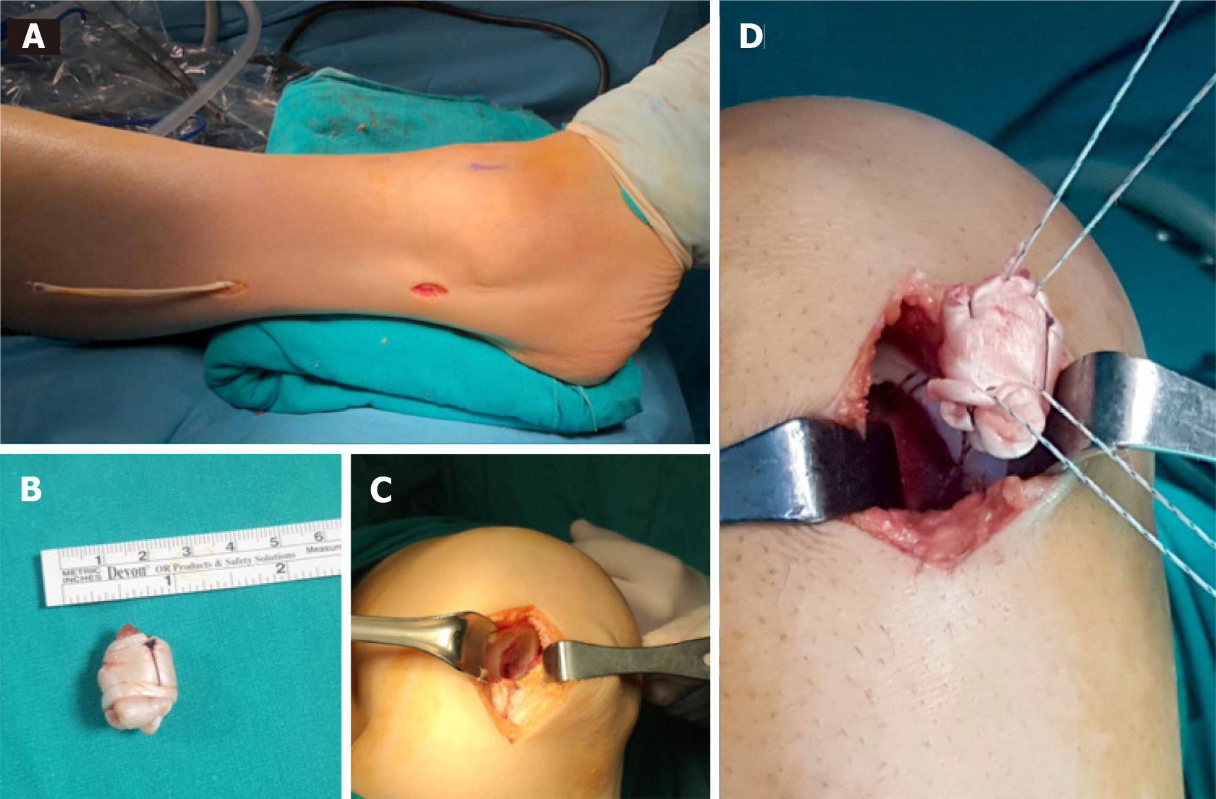
Figure 1 The technique of harvesting the autologous peroneal tendon from the donor area and placing it in the prepared osteochondral lesion area of the knee.
A: Two mini incisions were made in the ipsilateal leg to harvest a split portion of the peroneus longus tendon; B: Intaoperative image showing the rolled peroneus longus tendon with absorbable suture; C: Osteochondral defect was prepared for transplantation; D: The rolled tendon autograft was transplanted into the osteochondral defect space and fixed with suture anchor.
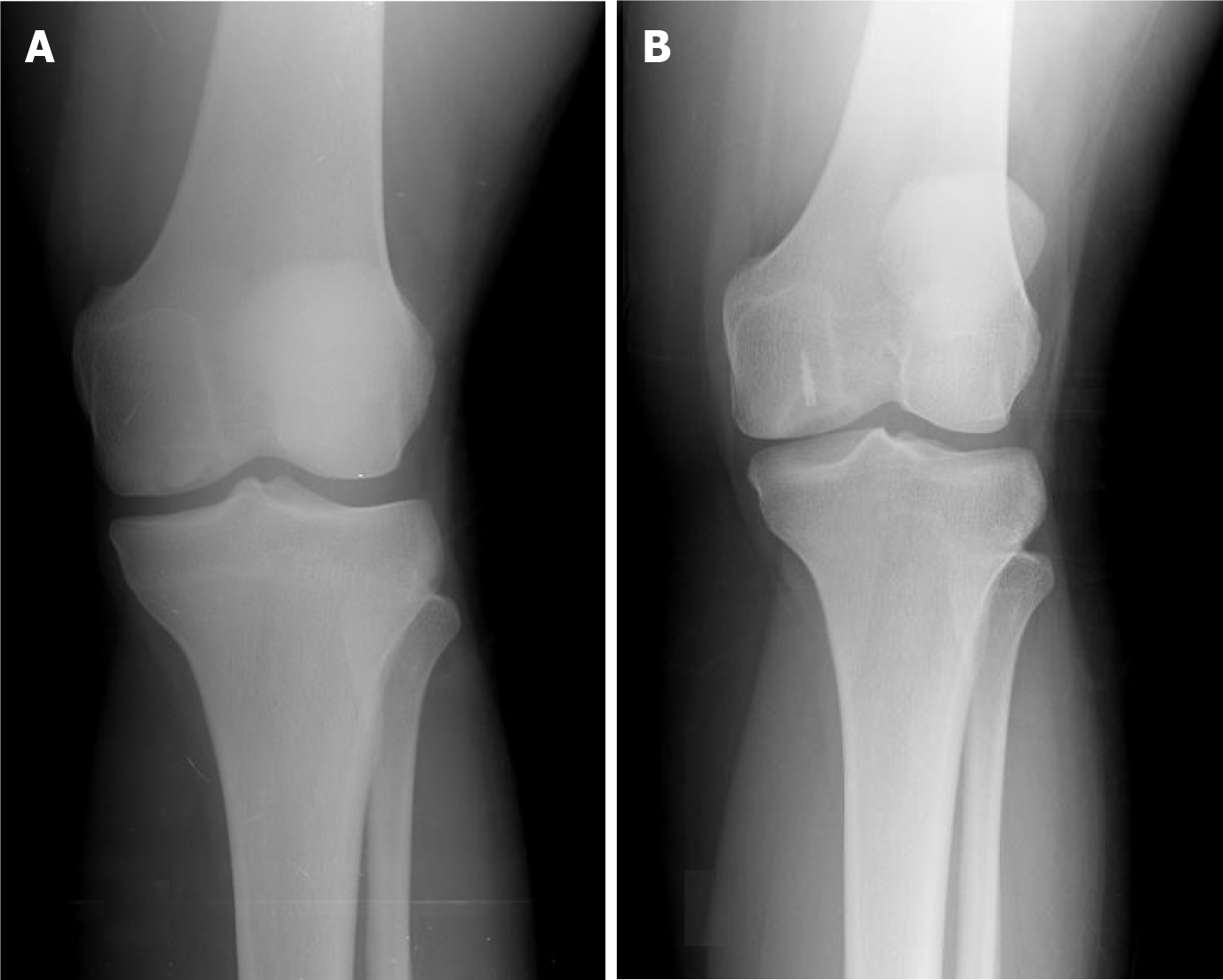
Figure 2 Radiographic images of a patient treated with the Autologous Tendon Transplantation technique.
A: Anteroposterior radiograph of the knee of a 24-year old female with a symptomatic lesion of the medial femoral condyle (4.6 cm2 size and 1.4 cm depth); B: Anteroposterior radiograph of the knee 6 years after autologous tendon transplantation with suture anchor show congruency of the articular surface of the medial femoral condyle. There are no arthritic changes. The patient is asymptomatic, free from pain and has no limitation of movement.
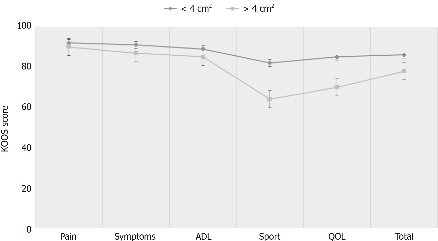
Figure 3 Knee injury and osteoarthritis outcome scores for different defect size.
Patients with less than 4 cm2 lesion had statistically significantly better overall knee injury and osteoarthritis outcome score than patients whose more than 4 cm2 lesion, particularly in sport and quality of life subscales. KOOS: Knee injury and osteoarthritis outcome score; ADL: Activity of daily living; QOL: Quality of life.
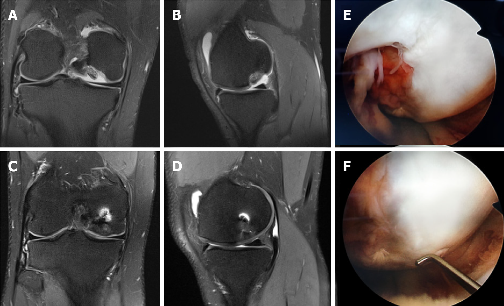
Figure 4 Magnetic resonance images and intraoperative images of a patient treated with the Autologous Tendon Transplantation technique.
A, B: Coronal and Sagittal T2 section magnetic resonance images (MRI) of a grade 4 osteochondral lesion involving articular surface of medial femoral condyle (3.4 cm2 size and 0.5 cm depth) in 24-year old male; C, D: 1-year follow-up coronal and sagittal T2 section MRI showing complete filling of the defect and establishment of smooth articular surface; E, F: Second look arthroscopy view at 1-year follow-up of grade 4 osteochondral lesion of medial femoral condyle showing filling of the defect with a well-integrated, smooth surfaced and stable regenerated cartilage.
- Citation: Turhan AU, Açıl S, Gül O, Öner K, Okutan AE, Ayas MS. Treatment of knee osteochondritis dissecans with autologous tendon transplantation: Clinical and radiological results. World J Orthop 2021; 12(11): 867-876
- URL: https://www.wjgnet.com/2218-5836/full/v12/i11/867.htm
- DOI: https://dx.doi.org/10.5312/wjo.v12.i11.867


