Copyright
©2013 Baishideng Publishing Group Co.
World J Clin Oncol. Aug 10, 2013; 4(3): 75-81
Published online Aug 10, 2013. doi: 10.5306/wjco.v4.i3.75
Published online Aug 10, 2013. doi: 10.5306/wjco.v4.i3.75
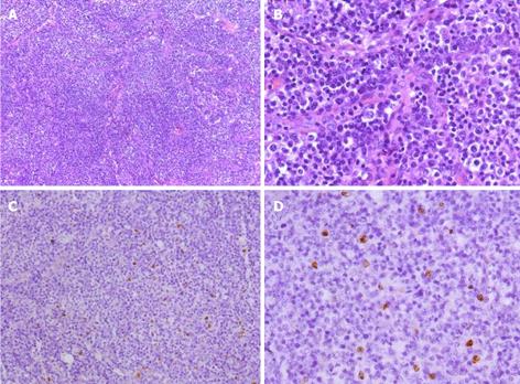
Figure 5 Photomicrograph of biopsy specimens in the celiac lymph nodes.
The normal architecture was lost, except for the presence of occasional depleted follicles with concentrically arranged follicular dendritic cells, and the architecture was effaced by polymorphic infiltrate with marked vascular proliferation. A weakly positive nuclear labeling of Epstein-Barr virus-encoded small nuclear RNA was observed in the celiac lymph nodes by in situ hybridization. A: Hematoxylin and eosin (H and E) staining, × 100; B: H and E staining, × 400. C: In situ hybridization staining for Epstein-Barr virus-encoded small nuclear RNA, × 100; D: In situ hybridization staining, × 400.
- Citation: Tao J, Zheng FP, Tian H, Lin Y, Li JZ, Chen XL, Chen JN, Shao CK, Wu B. Angioimmunoblastic T-cell lymphoma-associated pure red cell aplasia with abdominal pain. World J Clin Oncol 2013; 4(3): 75-81
- URL: https://www.wjgnet.com/2218-4333/full/v4/i3/75.htm
- DOI: https://dx.doi.org/10.5306/wjco.v4.i3.75


