Copyright
©2013 Baishideng Publishing Group Co.
World J Clin Oncol. Aug 10, 2013; 4(3): 75-81
Published online Aug 10, 2013. doi: 10.5306/wjco.v4.i3.75
Published online Aug 10, 2013. doi: 10.5306/wjco.v4.i3.75
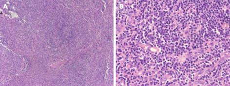
Figure 2 Photomicrograph of biopsy specimens of peripheral lymph nodes.
Significant inflammatory cell infiltration was observed; however, tumor cells, such as lymphoma cells, were not found in the specimens. A: Hematoxylin and eosin (HE) staining, × 100; B: HE staining, × 400.
- Citation: Tao J, Zheng FP, Tian H, Lin Y, Li JZ, Chen XL, Chen JN, Shao CK, Wu B. Angioimmunoblastic T-cell lymphoma-associated pure red cell aplasia with abdominal pain. World J Clin Oncol 2013; 4(3): 75-81
- URL: https://www.wjgnet.com/2218-4333/full/v4/i3/75.htm
- DOI: https://dx.doi.org/10.5306/wjco.v4.i3.75


