
















©2011 Baishideng Publishing Group Co.
World J Clin Oncol. May 10, 2011; 2(5): 229-236
Published online May 10, 2011. doi: 10.5306/wjco.v2.i5.229
Published online May 10, 2011. doi: 10.5306/wjco.v2.i5.229
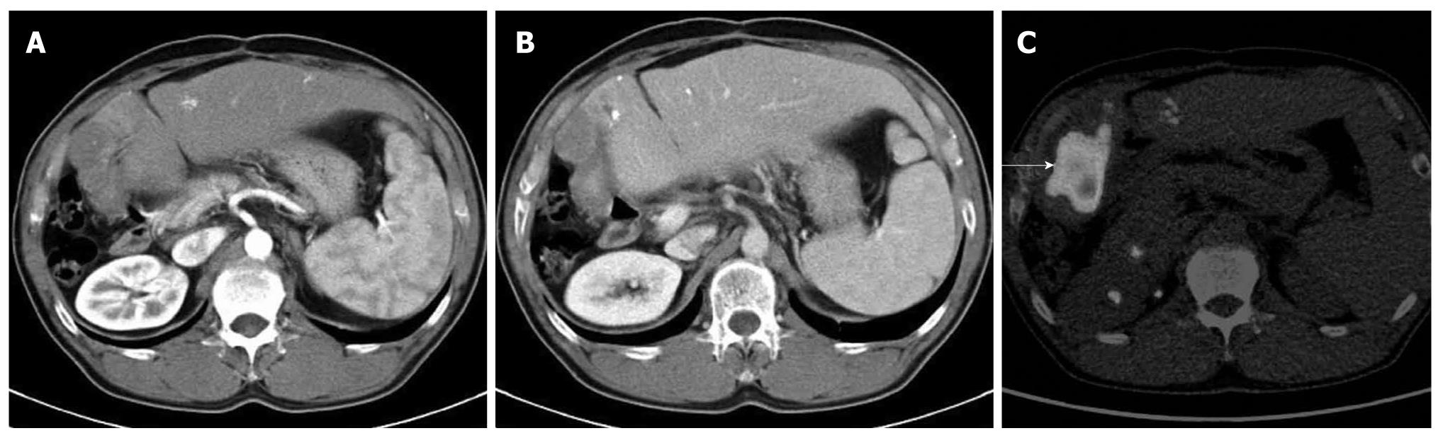
Figure 4 A case of recurrent hepatocellular carcinoma after right lobectomy.
A, B: Arterial and portal phase of dynamic computed tomography (CT). Although tumor shadow was noted at the cut surface of the liver, it is difficult to determine recurrence due to poor enhancement; C: The tumor was diagnosed as recurrence of hepatocellular carcinoma because of strong fluorodeoxyglucose deposit (arrows).
- Citation: Murakami K. FDG-PET for hepatobiliary and pancreatic cancer: Advances and current limitations. World J Clin Oncol 2011; 2(5): 229-236
- URL: https://www.wjgnet.com/2218-4333/full/v2/i5/229.htm
- DOI: https://dx.doi.org/10.5306/wjco.v2.i5.229