Copyright
©2011 Baishideng Publishing Group Co.
World J Clin Oncol. May 10, 2011; 2(5): 229-236
Published online May 10, 2011. doi: 10.5306/wjco.v2.i5.229
Published online May 10, 2011. doi: 10.5306/wjco.v2.i5.229
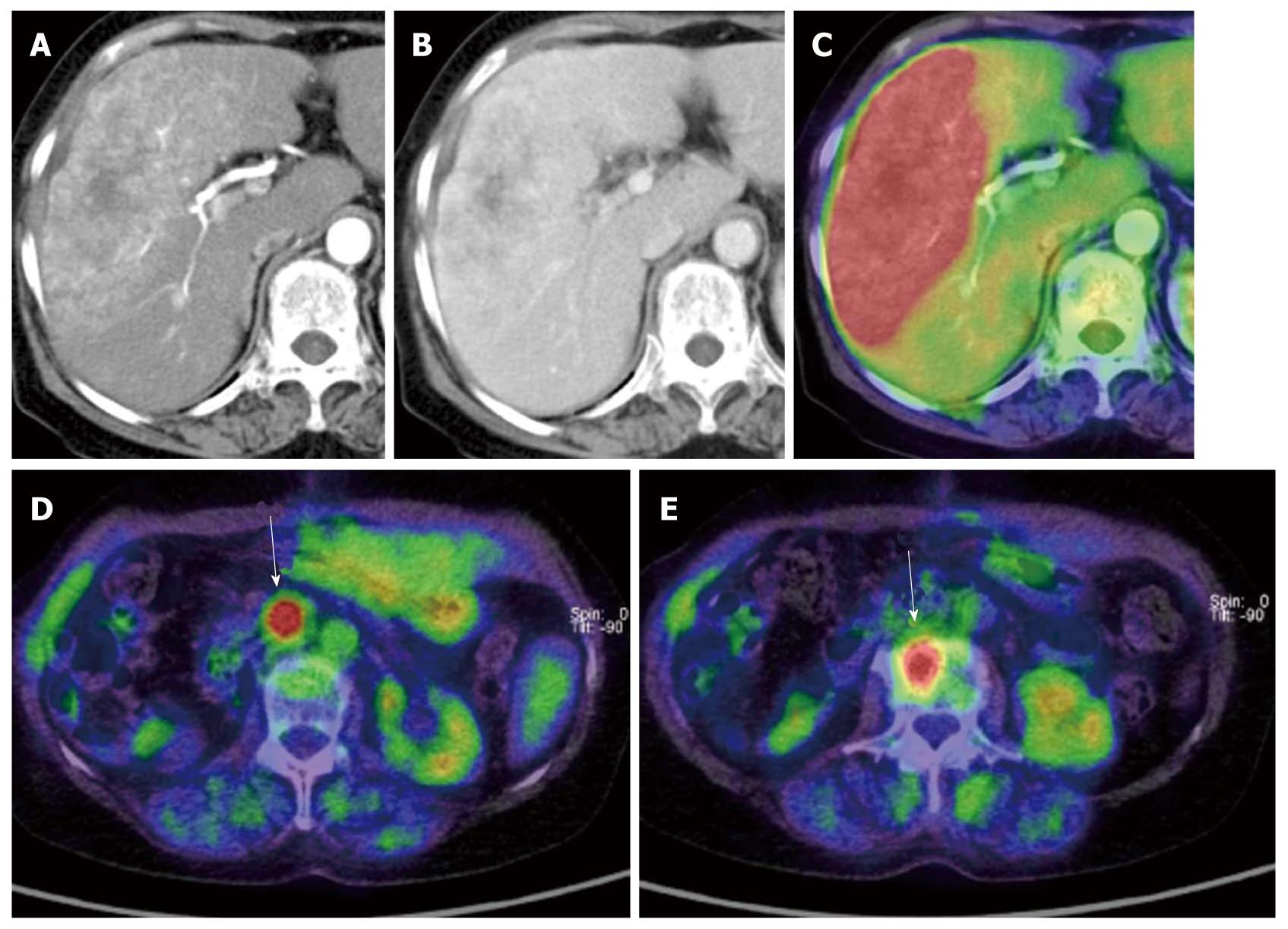
Figure 3 A case of mixed-type hepatocellular carcinoma with extrahepatic metastases.
A: Arterial phase of dynamic computed tomography (CT); B: Portal phase of dynamic CT. The tumor shows early enhancement which is a feature of hepatocellular carcinoma (HCC), although it also has a lobular border and delayed enhancement which are features of cholangiocellular carcinoma. Pathological diagnosis was mixed-type HCC; C: This type of HCC shows strong accumulation; D, E: This case also has lymph node and bone metastases (arrows).
- Citation: Murakami K. FDG-PET for hepatobiliary and pancreatic cancer: Advances and current limitations. World J Clin Oncol 2011; 2(5): 229-236
- URL: https://www.wjgnet.com/2218-4333/full/v2/i5/229.htm
- DOI: https://dx.doi.org/10.5306/wjco.v2.i5.229


