Copyright
©2011 Baishideng Publishing Group Co.
World J Clin Oncol. May 10, 2011; 2(5): 229-236
Published online May 10, 2011. doi: 10.5306/wjco.v2.i5.229
Published online May 10, 2011. doi: 10.5306/wjco.v2.i5.229
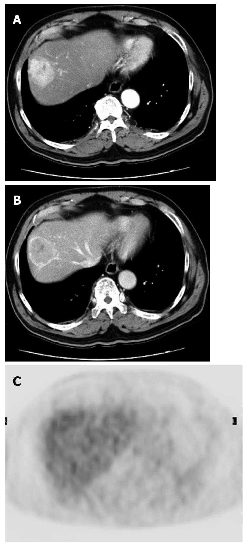
Figure 2 A case of well-differentiated hepatocellular carcinoma.
A, B: Arterial and portal phase of dynamic computed tomography. Typical enhancement pattern of hepatocellular carcinoma is shown; C: FDG-PET. There is no fluorodeoxyglucose uptake.
- Citation: Murakami K. FDG-PET for hepatobiliary and pancreatic cancer: Advances and current limitations. World J Clin Oncol 2011; 2(5): 229-236
- URL: https://www.wjgnet.com/2218-4333/full/v2/i5/229.htm
- DOI: https://dx.doi.org/10.5306/wjco.v2.i5.229


