Copyright
©2011 Baishideng Publishing Group Co.
World J Clin Oncol. Jan 10, 2011; 2(1): 8-27
Published online Jan 10, 2011. doi: 10.5306/wjco.v2.i1.8
Published online Jan 10, 2011. doi: 10.5306/wjco.v2.i1.8
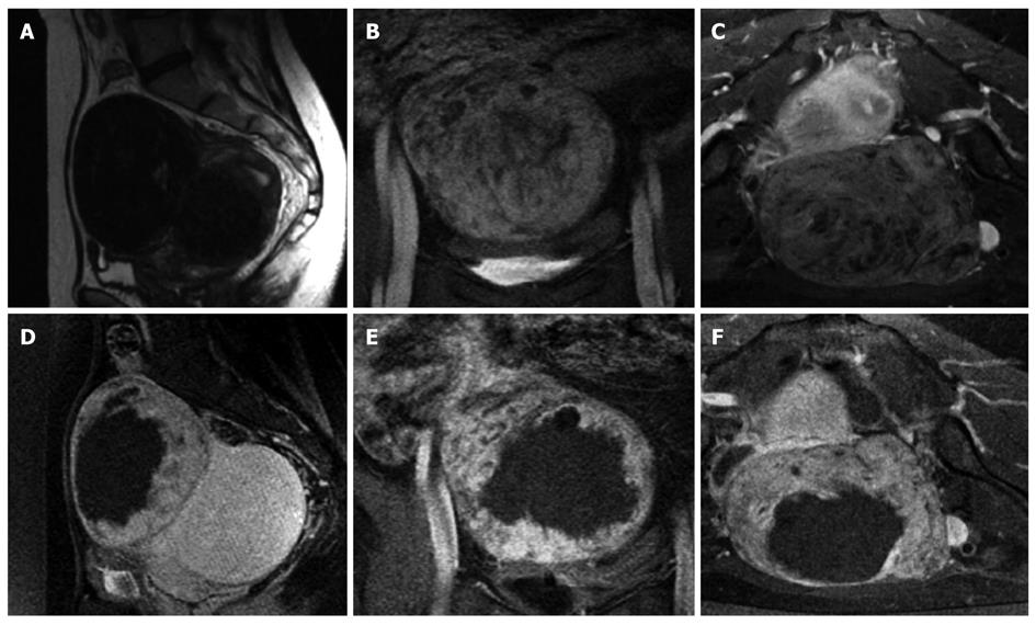
Figure 5 Images of a uterine fibroid pretreatment and posttreatment with MRgFUS.
Top, Sagittal T2 fast spin-echo (A), coronal spoiled gradient-recalled echo sequence (SPGR) postgadolinium (B), and axial SPGR postgadolinium (C) are obtained pretreatment. The low SI homogenous fibroid depicted in Figure 3A demonstrates slight heterogenous enhancement pretreatment (Figure 3B, C). Bottom, Sagittal SPGR post-gadolinium (D), coronal SPGR post-gadolinium (E), and axial SPGR post-gadolinium (F) are obtained immediately post-treatment. A new large nonperfused area is identified, consistent with treatment-induced necrosis.
- Citation: Zhou YF. High intensity focused ultrasound in clinical tumor ablation. World J Clin Oncol 2011; 2(1): 8-27
- URL: https://www.wjgnet.com/2218-4333/full/v2/i1/8.htm
- DOI: https://dx.doi.org/10.5306/wjco.v2.i1.8


