Copyright
©The Author(s) 2025.
World J Clin Oncol. Jun 24, 2025; 16(6): 105910
Published online Jun 24, 2025. doi: 10.5306/wjco.v16.i6.105910
Published online Jun 24, 2025. doi: 10.5306/wjco.v16.i6.105910
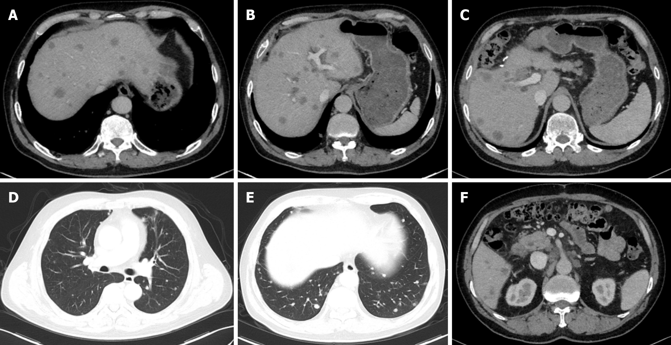
Figure 3 Computer tomography scans obtained on March 25, 2020, demonstrating extensive metastatic disease after surgery.
A-C: Diffuse liver metastases; D and E: Lung metastases; F: Retroperitoneal lymph nodes.
- Citation: Wang F, Gong XL, Chen XN, Yang ZH, Chu XY. Durable complete response to sintilimab plus chemotherapy in gallbladder cancer with extensive metastasis: A case report. World J Clin Oncol 2025; 16(6): 105910
- URL: https://www.wjgnet.com/2218-4333/full/v16/i6/105910.htm
- DOI: https://dx.doi.org/10.5306/wjco.v16.i6.105910


