Copyright
©The Author(s) 2025.
World J Clin Oncol. May 24, 2025; 16(5): 105341
Published online May 24, 2025. doi: 10.5306/wjco.v16.i5.105341
Published online May 24, 2025. doi: 10.5306/wjco.v16.i5.105341
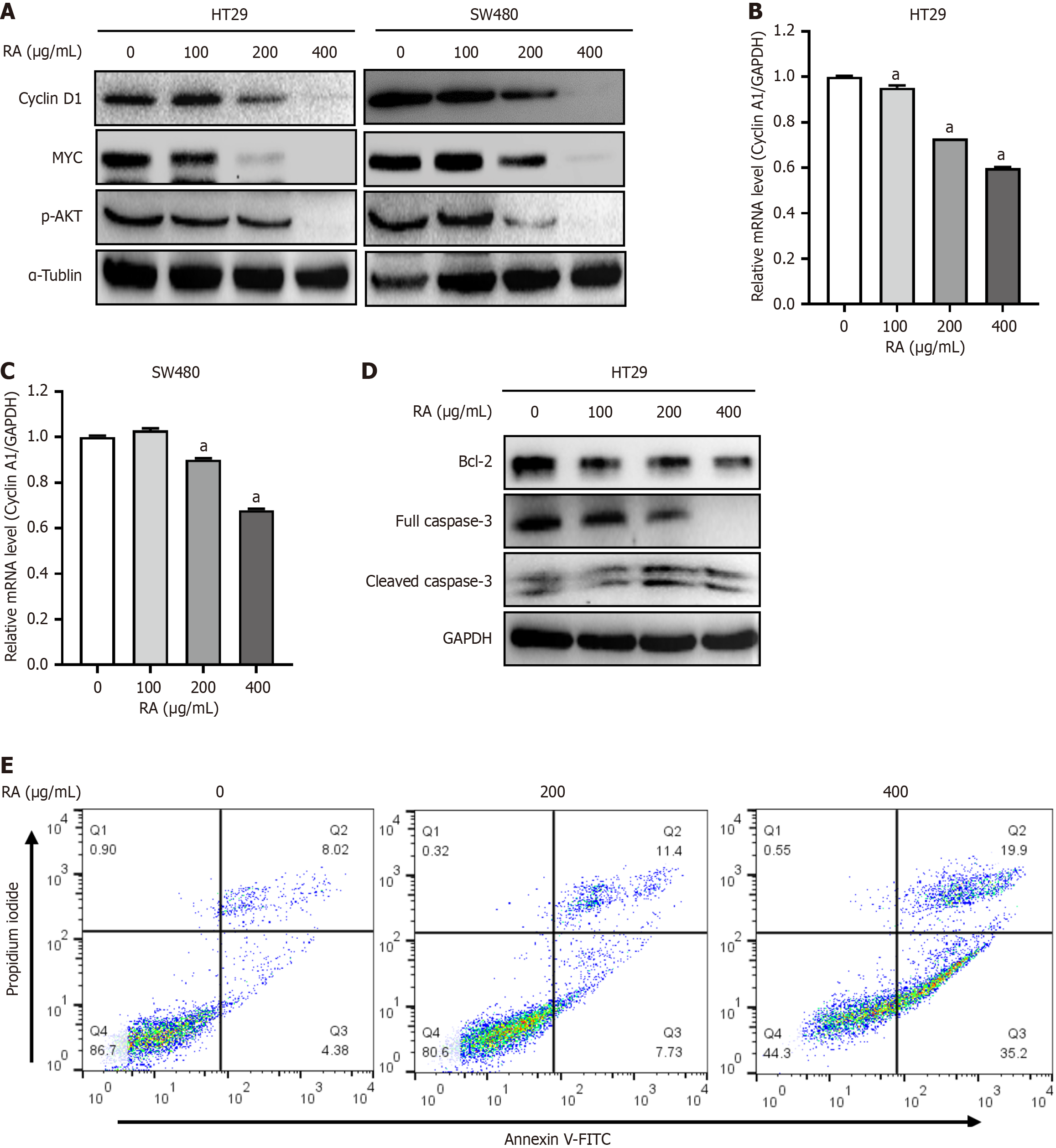
Figure 3 Rosmarinic acid inhibits the expressions of cell proliferative genes.
A: HT29 and SW480 cells were incubated with increasing concentrations of rosmarinic acid (RA) for 24 hours, followed by Western blot analysis using antibodies against cyclin D1 and MYC. α-Tublin was used as a loading control; B and C: The above cells were also prepared for quantitative real-time polymerase chain reaction to evaluate the mRNA levels of cyclin A1. GAPDH was used as an internal control; D: HT29 cells were treated with indicated concentrations of RA for 24 hours, followed by Western blot analysis using antibodies against Bcl-2, caspase-3, and GAPDH; E: HT29 cells were treated with indicated concentrations of RA for 24 hours, and then stained with Annexin V-FITC and propidium iodide. Percentages of Annexin V+ cells are indicated in the scatterplot (right low and upper quadrants). n = 3. Data are shown as the mean ± SD. aP < 0.0001 vs group ‘0’. RA: Rosmarinic acid; AKT: Protein kinase B.
- Citation: Liu WY, Wang H, Xu X, Wang X, Han KK, You WD, Yang Y, Zhang T. Natural compound rosmarinic acid displays anti-tumor activity in colorectal cancer cells by suppressing nuclear factor-kappa B signaling. World J Clin Oncol 2025; 16(5): 105341
- URL: https://www.wjgnet.com/2218-4333/full/v16/i5/105341.htm
- DOI: https://dx.doi.org/10.5306/wjco.v16.i5.105341


