Copyright
©The Author(s) 2025.
World J Clin Oncol. May 24, 2025; 16(5): 105341
Published online May 24, 2025. doi: 10.5306/wjco.v16.i5.105341
Published online May 24, 2025. doi: 10.5306/wjco.v16.i5.105341
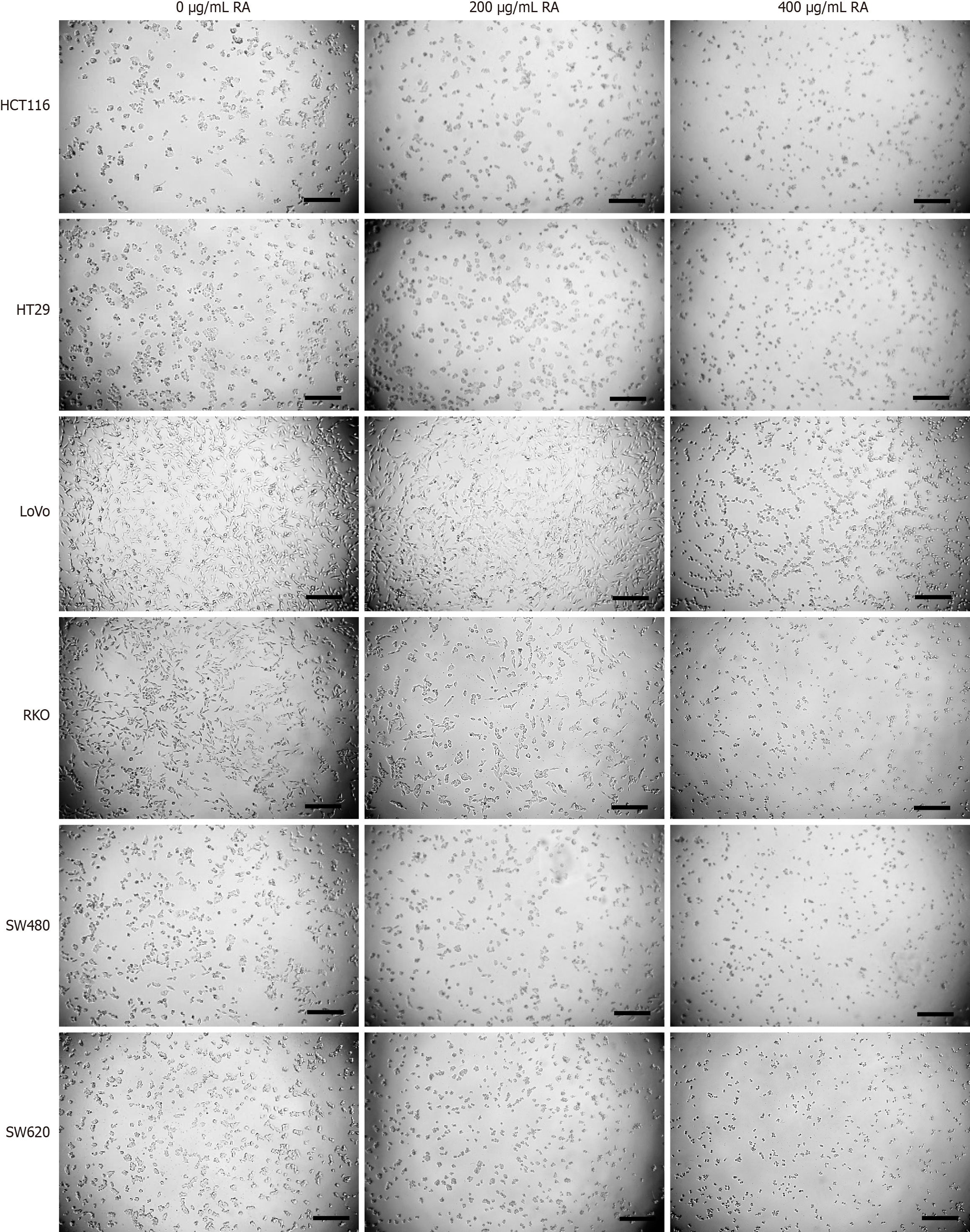
Figure 2 Optical microscopy images of colorectal cancer cells treated with rosmarinic acid.
Colorectal cancer cell lines, including HCT116, HT29, LoVo, RKO, SW480 and SW620, were separately incubated with increasing concentrations of rosmarinic acid. Twenty-four hours later, cells were taken photos under an optical microscopy. Scale bar = 0.3 mm. RA: Rosmarinic acid.
- Citation: Liu WY, Wang H, Xu X, Wang X, Han KK, You WD, Yang Y, Zhang T. Natural compound rosmarinic acid displays anti-tumor activity in colorectal cancer cells by suppressing nuclear factor-kappa B signaling. World J Clin Oncol 2025; 16(5): 105341
- URL: https://www.wjgnet.com/2218-4333/full/v16/i5/105341.htm
- DOI: https://dx.doi.org/10.5306/wjco.v16.i5.105341


