Copyright
©The Author(s) 2024.
World J Clin Oncol. Feb 24, 2024; 15(2): 178-194
Published online Feb 24, 2024. doi: 10.5306/wjco.v15.i2.178
Published online Feb 24, 2024. doi: 10.5306/wjco.v15.i2.178
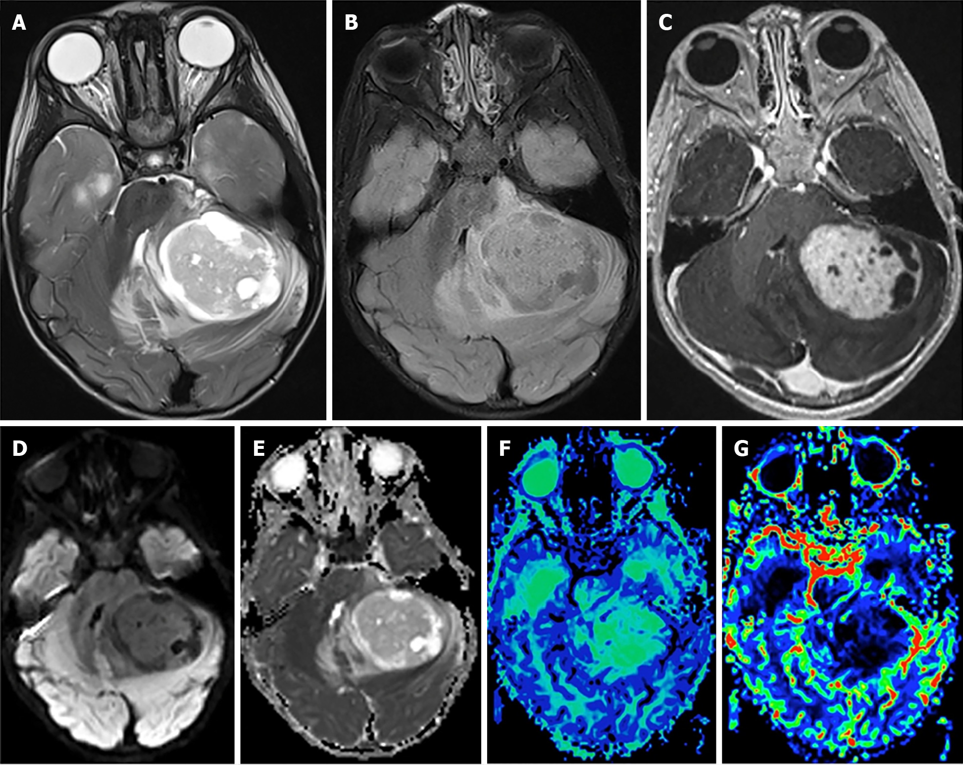
Figure 4 Pilocytic astrocytoma.
A: Axial T2-weighted scan through the cerebellum demonstrating a well-defined solid-cystic mass in the left cerebellar hemisphere that is heterogeneous onT2-weighted sequence and causes mass effect to the 4th ventricle. The solid component is minimally hyperintense compared to the gray matter; B: An axial T2 FLAIR image through the same level better demonstrates the area of T2 hyperintensity beyond the tumor margin, suggestive of edema; C: An axial post-contrast T1-weighted sequence through the same level demonstrates intense enhancement of the solid component of the tumor; D and E: The axial diffusion image and ADC map (E) through the same level show no diffusion restriction; F: The mean transit time map demonstrates increased transit time within the solid component of the tumor; G: The cerebral blood volume map shows that the tumor has very low cerebral blood volume. Citation: Bag AK, Chiang J, Patay Z. Radiohistogenomics of pediatric low-grade neuroepithelial tumors. Neuroradiology 2021; 63: 1185-1213. Copyright ©2021 The Authors. Published by Springer Nature[58] (Supplementary material).
- Citation: Mohamed AA, Alshaibi R, Faragalla S, Mohamed Y, Lucke-Wold B. Updates on management of gliomas in the molecular age. World J Clin Oncol 2024; 15(2): 178-194
- URL: https://www.wjgnet.com/2218-4333/full/v15/i2/178.htm
- DOI: https://dx.doi.org/10.5306/wjco.v15.i2.178


