Copyright
©The Author(s) 2017.
World J Gastrointest Pharmacol Ther. Feb 6, 2017; 8(1): 47-59
Published online Feb 6, 2017. doi: 10.4292/wjgpt.v8.i1.47
Published online Feb 6, 2017. doi: 10.4292/wjgpt.v8.i1.47
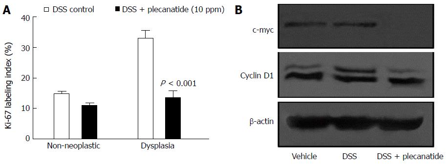
Figure 4 Effect of plecanatide on markers of proliferation in colonic epithelial cells from Apc+/Min-FCCC mice with dextran sodium sulfate-induced inflammation.
A: Normal (non-neoplastic) and neoplastic colon tissues from mice treated with DSS only and DSS + plecanatide (10 ppm) were stained with antibodies specific for Ki-67. Nuclear staining of Ki-67 (positive) was recorded as a labeling index (number of positive cells/total number of cells evaluated; mean ± SEM). Statistical comparisons between DSS control and DSS plus plecanatide-treated groups (n = 7-9 mice per group) were performed using the Student t test. A P value ≤ 0.05 was considered significant; B: Colon tissue (1 cm) from 6 animals per group was pooled to prepare cell lysates. A representative immunoblot demonstrating the effect of plecanatide on expression of c-Myc and cyclin D1 is shown. β-actin was used to normalize protein loading. DSS: Dextran sodium sulfate.
- Citation: Chang WCL, Masih S, Thadi A, Patwa V, Joshi A, Cooper HS, Palejwala VA, Clapper ML, Shailubhai K. Plecanatide-mediated activation of guanylate cyclase-C suppresses inflammation-induced colorectal carcinogenesis in Apc+/Min-FCCC mice. World J Gastrointest Pharmacol Ther 2017; 8(1): 47-59
- URL: https://www.wjgnet.com/2150-5349/full/v8/i1/47.htm
- DOI: https://dx.doi.org/10.4292/wjgpt.v8.i1.47


