Copyright
©The Author(s) 2020.
World J Gastrointest Pharmacol Ther. Nov 8, 2020; 11(5): 93-109
Published online Nov 8, 2020. doi: 10.4292/wjgpt.v11.i5.93
Published online Nov 8, 2020. doi: 10.4292/wjgpt.v11.i5.93
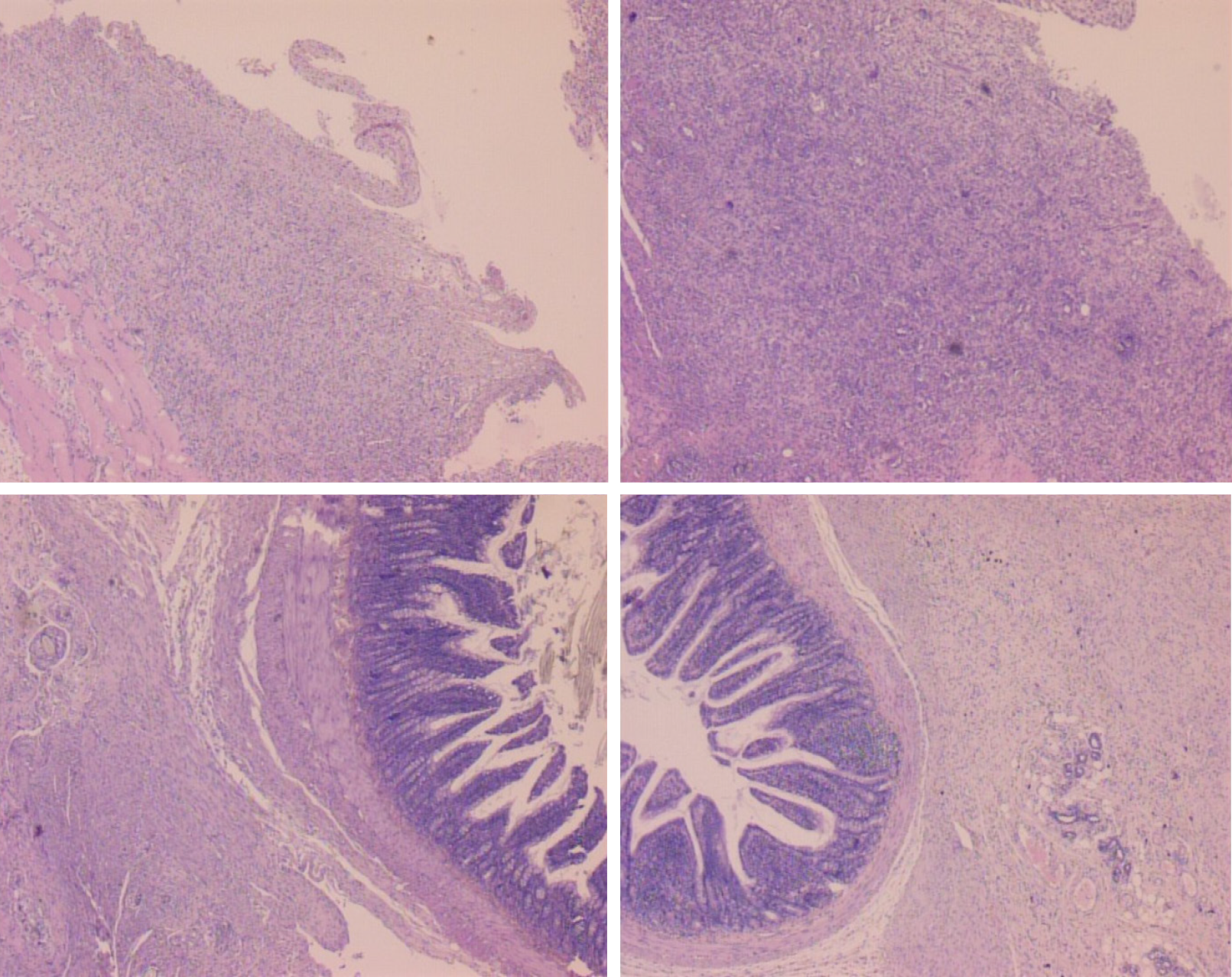
Figure 5 Histological assessment after 7 d (upper) and 14 d (low), in rats subjected to the excision of the parietal peritoneum with underlying superficial layer of muscle tissue, controls [upper left (7 d), low left (14 )] and BPC 157-treated rats [upper right (7 d), low right (14 d)].
7 d. Upper, left, controls. Edematous, relatively poorly formed granulation tissue, covering large areas of serosa. Upper, right, BPC 157. By far smaller areas covered with more dense and mature granulation tissue with vessels appearing more mature and better formed. 14 d. Low, left, controls. After two weeks large areas of still poorly organized granulation tissue invading the bowel and abdominal wall. Low, right, BPC 157. Young connective tissue scar is formed in smaller areas, with poor invasion into the bowel serosa/subserosa leading to very limited and non-strangulating adhesions. HE stain, objective × 10.
- Citation: Berkopic Cesar L, Gojkovic S, Krezic I, Malekinusic D, Zizek H, Batelja Vuletic L, Petrovic A, Horvat Pavlov K, Drmic D, Kokot A, Vlainic J, Seiwerth S, Sikiric P. Bowel adhesion and therapy with the stable gastric pentadecapeptide BPC 157, L-NAME and L-arginine in rats. World J Gastrointest Pharmacol Ther 2020; 11(5): 93-109
- URL: https://www.wjgnet.com/2150-5349/full/v11/i5/93.htm
- DOI: https://dx.doi.org/10.4292/wjgpt.v11.i5.93


