
















©The Author(s) 2018.
World J Gastrointest Pathophysiol. Oct 25, 2018; 9(3): 63-72
Published online Oct 25, 2018. doi: 10.4291/wjgp.v9.i3.63
Published online Oct 25, 2018. doi: 10.4291/wjgp.v9.i3.63
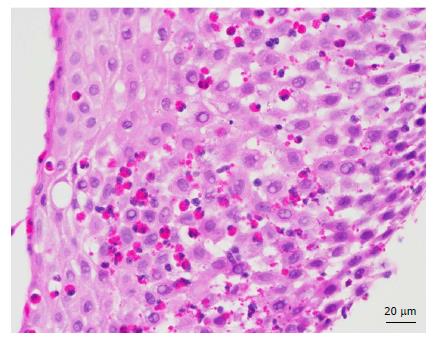
Figure 3 Histological specimen from the oesophagus (luminal aspect on left) of an eosinophilic oesophagitis patient showing marked oedema and numerous intraepithelial eosinophils in the oesophageal squamous mucosa, which are also seen in the superficial component of the mucosa.
- Citation: Wong S, Ruszkiewicz A, Holloway RH, Nguyen NQ. Gastro-oesophageal reflux disease and eosinophilic oesophagitis: What is the relationship? World J Gastrointest Pathophysiol 2018; 9(3): 63-72
- URL: https://www.wjgnet.com/2150-5330/full/v9/i3/63.htm
- DOI: https://dx.doi.org/10.4291/wjgp.v9.i3.63