Copyright
©The Author(s) 2017.
World J Gastrointest Pathophysiol. Nov 15, 2017; 8(4): 150-160
Published online Nov 15, 2017. doi: 10.4291/wjgp.v8.i4.150
Published online Nov 15, 2017. doi: 10.4291/wjgp.v8.i4.150
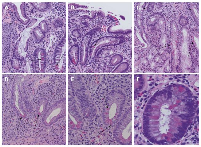
Figure 3 Examples of Paneth cell metaplasia throughout the intestinal tract.
A: Paneth cell metaplasia (arrows) in Barrett mucosa. Squamous epithelia of the oesophagus are marked with an arrowhead; B: Chronic atrophic gastritis with Paneth cell metaplasia (arrow); C: Paneth cell metaplasia (arrows) of Brunner’s gland. In the upper part, small intestinal mucosa and secretory ducts are shown (arrowheads); D: Colon mucosa in ulcerative colitis with disturbed crypt architecture, increased numbers of stroma infiltrating inflammatory cells, and Paneth cell metaplasia (arrowheads); E: Higher magnification of ulcerative colitis-associated Paneth cell metaplasia (arrows) in colon mucosa as demonstrated in (D); F: Paneth cell metaplasia (arrows) in tubular adenoma of the colon with low-grade dysplasia.
- Citation: Gassler N. Paneth cells in intestinal physiology and pathophysiology. World J Gastrointest Pathophysiol 2017; 8(4): 150-160
- URL: https://www.wjgnet.com/2150-5330/full/v8/i4/150.htm
- DOI: https://dx.doi.org/10.4291/wjgp.v8.i4.150


