Copyright
©The Author(s) 2017.
World J Gastrointest Pathophysiol. Aug 15, 2017; 8(3): 142-149
Published online Aug 15, 2017. doi: 10.4291/wjgp.v8.i3.142
Published online Aug 15, 2017. doi: 10.4291/wjgp.v8.i3.142
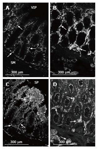
Figure 5 VIP and SP nerve fibres in the mucosa in normal colon and MEN2B colon.
VIP-immunoreactivity in (A) Control (HSCR) and (B) MEN2B patient. Crypts in transverse colon. VIP immunoreactive nerve fibres (arrows) are plentiful at the base and within the arms of the crypts in the control. There is a large increase in the numbers of VIP-IR nerve fibres and brightness of labelling in the crypts. SP-immunoreactivity in (C) Control (HSCR) and (D) MEN2B patient. Background labelling is higher with SP with labelling of epithelial cells. Varicose nerve fibres are visible (arrows) in a similar pattern to VIP. The thickness and brightness of the nerve fibres is greatly increased within the crypts and in the submucosa in MEN2B. Note all images are at the same magnification.
- Citation: Hutson JM, Farmer PJ, Peck CJ, Chow CW, Southwell BR. Multiple endocrine neoplasia 2B: Differential increase in enteric nerve subgroups in muscle and mucosa. World J Gastrointest Pathophysiol 2017; 8(3): 142-149
- URL: https://www.wjgnet.com/2150-5330/full/v8/i3/142.htm
- DOI: https://dx.doi.org/10.4291/wjgp.v8.i3.142


