Copyright
©The Author(s) 2017.
World J Gastrointest Pathophysiol. May 15, 2017; 8(2): 27-38
Published online May 15, 2017. doi: 10.4291/wjgp.v8.i2.27
Published online May 15, 2017. doi: 10.4291/wjgp.v8.i2.27
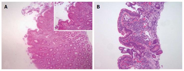
Figure 2 Histological features of celiac disease.
A: Example of tissue scored as Marsh 2 characterized by lymphocytic enteritis with crypt hyperplasia: Intraepithelial lymphocytosis and elongation and branching of crypts in which there is an increased proliferation of epithelial cells; B: Example of tissue scored as Marsh 3A characterized by partial villous atrophy, the villi are blunt and shortened. Arbitrarily, samples are classified as partial villous atrophy if the villus-crypt ratio was less than 1:1 (Objective magnification × 4, inset × 10).
- Citation: Parzanese I, Qehajaj D, Patrinicola F, Aralica M, Chiriva-Internati M, Stifter S, Elli L, Grizzi F. Celiac disease: From pathophysiology to treatment. World J Gastrointest Pathophysiol 2017; 8(2): 27-38
- URL: https://www.wjgnet.com/2150-5330/full/v8/i2/27.htm
- DOI: https://dx.doi.org/10.4291/wjgp.v8.i2.27


