Copyright
©The Author(s) 2015.
World J Gastrointest Pathophysiol. Nov 15, 2015; 6(4): 140-149
Published online Nov 15, 2015. doi: 10.4291/wjgp.v6.i4.140
Published online Nov 15, 2015. doi: 10.4291/wjgp.v6.i4.140
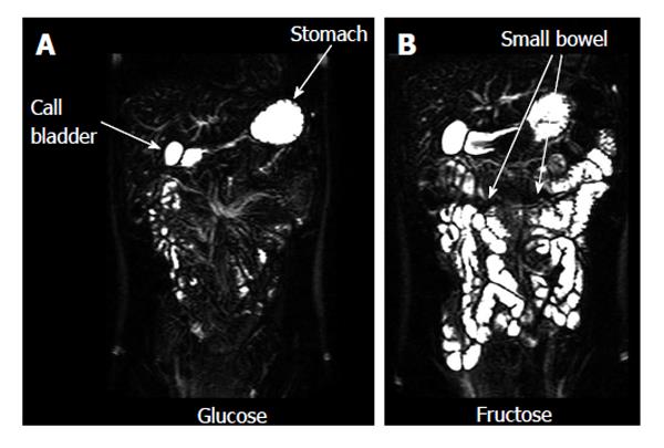
Figure 4 Small bowel water imaging.
Representative example of coronal images of the small bowel water from a single volunteer acquired 75 min after drinking 40 g of glucose (A) or 40 g fructose (B) in 500 mL water. Glucose is rapidly absorbed so the small bowel has very little water in it despite the large drink. Conversely fructose is poorly absorbed and osmotically active as shown by the large amount of water in the small bowel. Adapted with authors’ own copyright from ref. [77].
- Citation: Khalaf A, Hoad CL, Spiller RC, Gowland PA, Moran GW, Marciani L. Magnetic resonance imaging biomarkers of gastrointestinal motor function and fluid distribution. World J Gastrointest Pathophysiol 2015; 6(4): 140-149
- URL: https://www.wjgnet.com/2150-5330/full/v6/i4/140.htm
- DOI: https://dx.doi.org/10.4291/wjgp.v6.i4.140


