Copyright
©2014 Baishideng Publishing Group Inc.
World J Gastrointest Pathophysiol. Nov 15, 2014; 5(4): 487-495
Published online Nov 15, 2014. doi: 10.4291/wjgp.v5.i4.487
Published online Nov 15, 2014. doi: 10.4291/wjgp.v5.i4.487
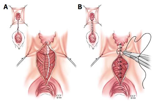
Figure 3 Episioproctotomy.
A: Episioproctotomy begins with fistulotomy and division of all tissue overlying the fistula, including sphincter muscles and rectal and vaginal walls. Complete debridement of the granulation tissue of the fistula tract is carried out along with the lateral identification and mobilization of the sphincter muscles; B: The rectal mucosa is repaired followed by an overlap repair of the sphincter muscles. The repair is completed by closing the vaginal mucosa. Reprinted with permission, Cleveland Clinic Center for Medical Art and Photography © 1999-2014. All Rights Reserved.
- Citation: Valente MA, Hull TL. Contemporary surgical management of rectovaginal fistula in Crohn's disease. World J Gastrointest Pathophysiol 2014; 5(4): 487-495
- URL: https://www.wjgnet.com/2150-5330/full/v5/i4/487.htm
- DOI: https://dx.doi.org/10.4291/wjgp.v5.i4.487


