Copyright
©2014 Baishideng Publishing Group Inc.
World J Gastrointest Pathophysiol. Aug 15, 2014; 5(3): 252-270
Published online Aug 15, 2014. doi: 10.4291/wjgp.v5.i3.252
Published online Aug 15, 2014. doi: 10.4291/wjgp.v5.i3.252
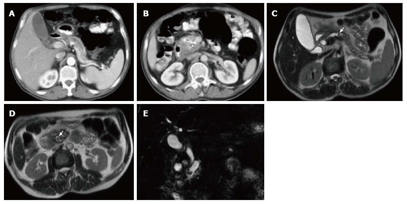
Figure 17 Chronic pancreatitis with pancreatic parenchymal calcifications and pancreatic duct stones.
A, B: Axial CT scan during the late arterial phase; C, D: Axial T2 single-shot fast spin-echo images; E: Coronal 3D- Cholangiopancreatogram (MRCP) image. CT shows a markedly dilated and tortuous main pancreatic duct (MPD) (A, B), with foci of thick calcification involving the pancreatic head and uncinate process parenchyma (B). Large the proximal MPD stone was suspected on CT (arrow, B). MRCP shows gross pancreatic ductal dilatation with confirmation of the distal intraductal calculus (arrow, D), and shows an additional mid-pancreatic duct stone, not clearly seen on CT (arrow, C). No pancreatic masses or ductal anomalies are identified.
- Citation: Busireddy KK, AlObaidy M, Ramalho M, Kalubowila J, Baodong L, Santagostino I, Semelka RC. Pancreatitis-imaging approach. World J Gastrointest Pathophysiol 2014; 5(3): 252-270
- URL: https://www.wjgnet.com/2150-5330/full/v5/i3/252.htm
- DOI: https://dx.doi.org/10.4291/wjgp.v5.i3.252


