Copyright
©2014 Baishideng Publishing Group Inc.
World J Gastrointest Pathophysiol. Aug 15, 2014; 5(3): 252-270
Published online Aug 15, 2014. doi: 10.4291/wjgp.v5.i3.252
Published online Aug 15, 2014. doi: 10.4291/wjgp.v5.i3.252
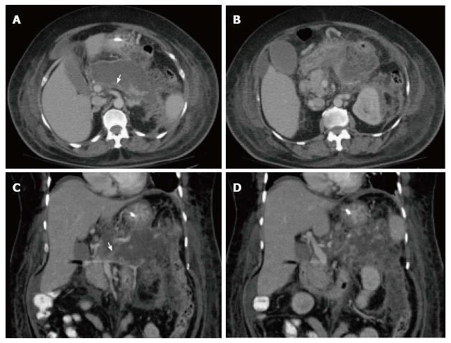
Figure 7 Severe acute necrotizing pancreatitis and peri pancreatitis.
A-B: Axial CT scan during the late arterial phase; C-D: Coronal reformatted CT images. There is evidence of lack of arterial enhancement involving the pancreatic body and tail, which are replaced by necrotic tissue, associated with heterogenous peripancreatic tissue inflammation and necrosis extending to left perinephric space (A-B) and paracolic gutter (C-D), in keeping with severe necrotizing pancreatitis and peripancreatitis. There is also evidence of splenic vein thrombosis (arrow, A, C), a known complication of acute pancreatitis.
- Citation: Busireddy KK, AlObaidy M, Ramalho M, Kalubowila J, Baodong L, Santagostino I, Semelka RC. Pancreatitis-imaging approach. World J Gastrointest Pathophysiol 2014; 5(3): 252-270
- URL: https://www.wjgnet.com/2150-5330/full/v5/i3/252.htm
- DOI: https://dx.doi.org/10.4291/wjgp.v5.i3.252


