
















©2013 Baishideng Publishing Group Co.
World J Gastrointest Pathophysiol. Aug 15, 2013; 4(3): 53-58
Published online Aug 15, 2013. doi: 10.4291/wjgp.v4.i3.53
Published online Aug 15, 2013. doi: 10.4291/wjgp.v4.i3.53
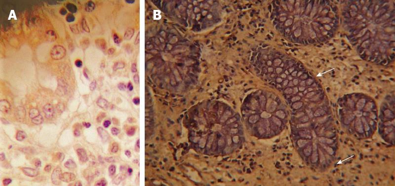
Figure 2 Immunohistochemical staining of syndecan 1 in the lamina propria in controls (A) and Crohn’s disease (B) stenotic complication.
In normal tissue staining is confined to basolateral area of the crypts (arrow). In Crohn’s disease, positive stromal cells and apical epithelium are observed stained brown (diaminobenzidine). Negative cells are counterstained blue (haematoxylin) (× 400).
- Citation: Ierardi E, Giorgio F, Piscitelli D, Principi M, Cantatore S, Fiore MG, Rossi R, Barone M, Di Leo A, Panella C. Altered molecular pattern of mucosal healing in Crohn’s disease fibrotic stenosis. World J Gastrointest Pathophysiol 2013; 4(3): 53-58
- URL: https://www.wjgnet.com/2150-5330/full/v4/i3/53.htm
- DOI: https://dx.doi.org/10.4291/wjgp.v4.i3.53