Copyright
©2012 Baishideng Publishing Group Co.
World J Gastrointest Pathophysiol. Dec 15, 2012; 3(6): 102-108
Published online Dec 15, 2012. doi: 10.4291/wjgp.v3.i6.102
Published online Dec 15, 2012. doi: 10.4291/wjgp.v3.i6.102
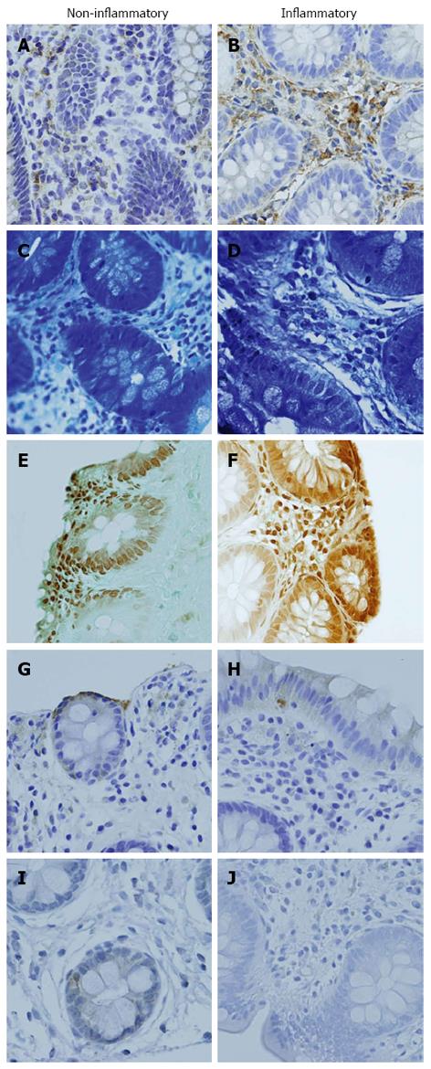
Figure 2 Immunohistochemical staining (40 ×).
Examples of labeling of interleukin-6 (A-B), mast cells (C-D), enterochromaffin cells cells (E-F), 5-hydroxytryptamine (G-H) and substance P (I-J) in non-inflammatory and inflammatory biopsies are shown.
- Citation: Henderson WA, Shankar R, Taylor TJ, Del Valle-Pinero AY, Kleiner DE, Kim KH, Youssef NN. Inverse relationship of interleukin-6 and mast cells in children with inflammatory and non-inflammatory abdominal pain phenotypes. World J Gastrointest Pathophysiol 2012; 3(6): 102-108
- URL: https://www.wjgnet.com/2150-5330/full/v3/i6/102.htm
- DOI: https://dx.doi.org/10.4291/wjgp.v3.i6.102


