
















©The Author(s) 2025.
World J Gastrointest Pathophysiol. Dec 22, 2025; 16(4): 112822
Published online Dec 22, 2025. doi: 10.4291/wjgp.v16.i4.112822
Published online Dec 22, 2025. doi: 10.4291/wjgp.v16.i4.112822
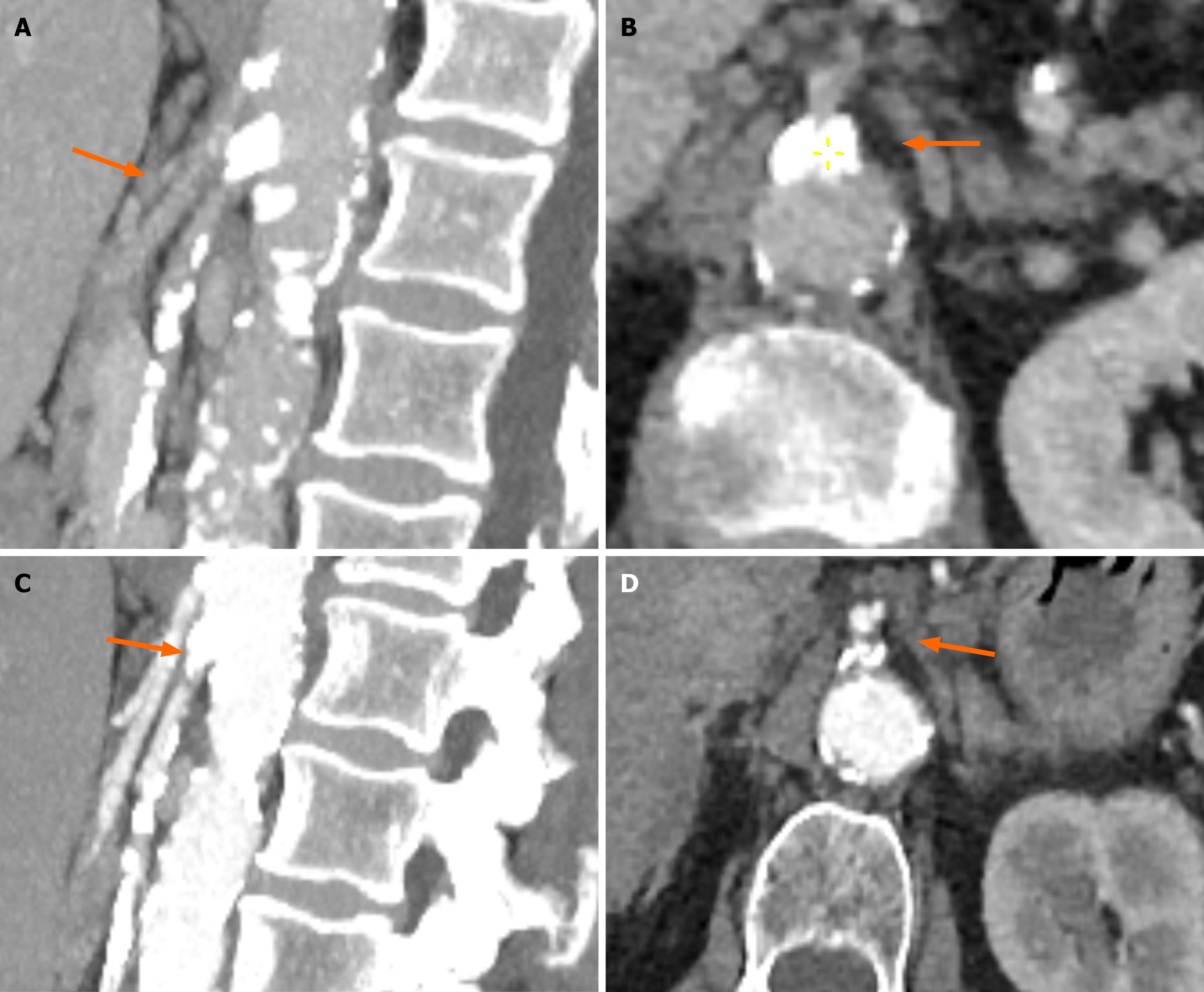
Figure 2 Computed tomography abdomen protocol and angiography protocol.
A and B: Show the presence of significant calcification at both the celiac trunk and superior mesenteric artery (SMA) (A: Sagittal reconstruction), with extensive calcification at the SMA ostium when viewed in axial view. The SMA show extensive calcification along the entirety of its length especially in sagittal view; C and D: Show extensive calcifications at both the celiac trunk and SMA (C: Sagittal reconstruction), extensive calcification at the SMA ostium when viewed in axial view (D).
- Citation: Abdulrasak M, Wernehov I, Holmgren J. Chronic mesenteric ischemia diagnosed via incidental CT findings with gastroenterologist perspective: Two case reports. World J Gastrointest Pathophysiol 2025; 16(4): 112822
- URL: https://www.wjgnet.com/2150-5330/full/v16/i4/112822.htm
- DOI: https://dx.doi.org/10.4291/wjgp.v16.i4.112822