
















©The Author(s) 2025.
World J Gastrointest Pathophysiol. Dec 22, 2025; 16(4): 112695
Published online Dec 22, 2025. doi: 10.4291/wjgp.v16.i4.112695
Published online Dec 22, 2025. doi: 10.4291/wjgp.v16.i4.112695
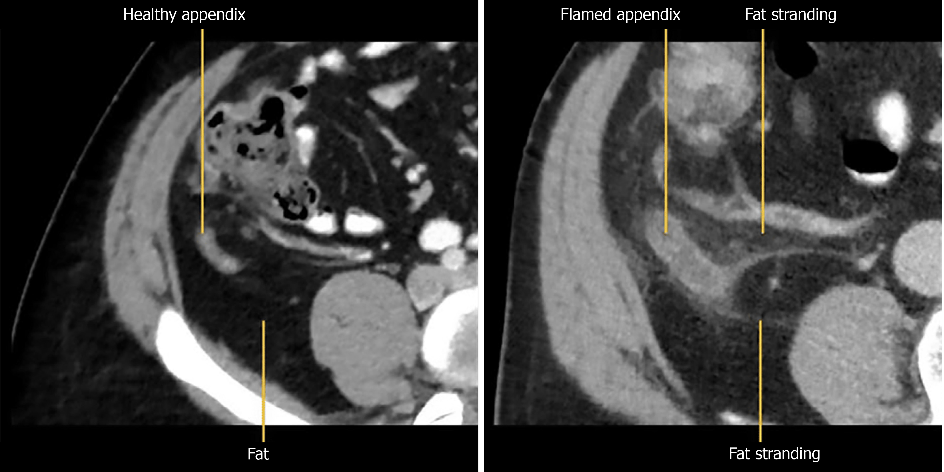
Figure 1 On the left is a computed tomography image of normal healthy appendix while on the right is an inflamed appendix with fat stranding.
- Citation: Perera Molligoda Arachchige AS, Teixeira de Castro Gonçalves Ortega AC, Amorim Moreira Alves G. Imaging of appendicitis: Computed tomography, magnetic resonance imaging, and ultrasound in diagnosis and management. World J Gastrointest Pathophysiol 2025; 16(4): 112695
- URL: https://www.wjgnet.com/2150-5330/full/v16/i4/112695.htm
- DOI: https://dx.doi.org/10.4291/wjgp.v16.i4.112695