
















©The Author(s) 2025.
World J Gastrointest Pathophysiol. Sep 22, 2025; 16(3): 107273
Published online Sep 22, 2025. doi: 10.4291/wjgp.v16.i3.107273
Published online Sep 22, 2025. doi: 10.4291/wjgp.v16.i3.107273
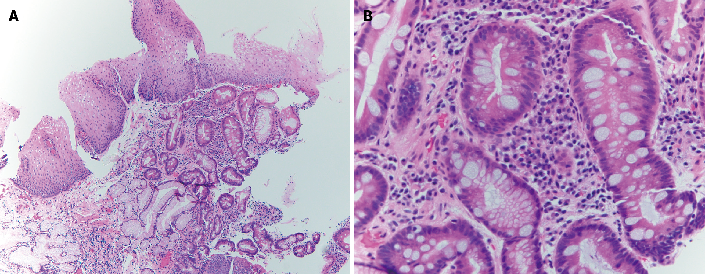
Figure 3 Pathology images showing intestinal metaplasia.
A: Hematoxylin and eosin stain (magnification 10 ×) of esophageal biopsy showing squamous epithelium overlying the glands with intestinal metaplasia; B: Hematoxylin and eosin stain (magnification 40 ×) of esophageal biopsy showing columnar epithelium with intestinal metaplasia characterized by barrel shaped goblet cells filled with bluish cytoplasmic hue (mucin-rich).
- Citation: Chela HK, Gandhi M, Hazique M, Ertugrul H, Gangu K, Daglilar E. Barrett’s esophagus: A review. World J Gastrointest Pathophysiol 2025; 16(3): 107273
- URL: https://www.wjgnet.com/2150-5330/full/v16/i3/107273.htm
- DOI: https://dx.doi.org/10.4291/wjgp.v16.i3.107273