
















©The Author(s) 2025.
World J Gastrointest Pathophysiol. Sep 22, 2025; 16(3): 106014
Published online Sep 22, 2025. doi: 10.4291/wjgp.v16.i3.106014
Published online Sep 22, 2025. doi: 10.4291/wjgp.v16.i3.106014
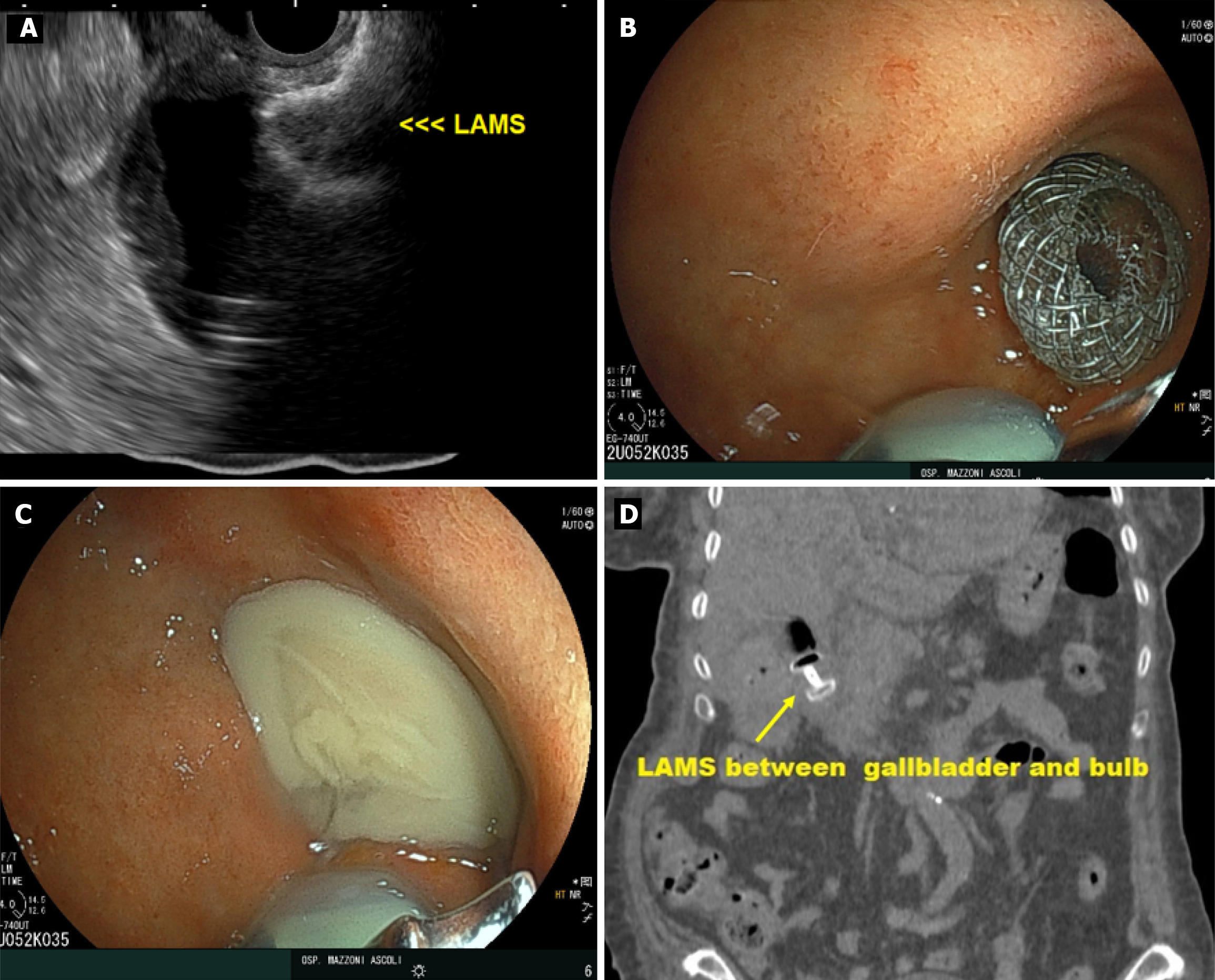
Figure 2 Endoscopic ultrasound-guided gallbladder drainage.
A: Endoscopic ultrasound view of the lumen-apposing metal stent (LAMS) inside the gallbladder; B: LAMS placed through the duodenal bulb; C: Pus coming from the gallbladder; D: CT image of the LAMS between the gallbladder and bulb. LAMS: Lumen-apposing metal stent.
- Citation: Antonini F, Donnarumma D, Buono T, Di Saverio S, Gardini A. Single-session endoscopic ultrasound-guided gallbladder drainage and biopsy in pancreatic cancer, obstructive jaundice, and acute cholecystitis: A case report. World J Gastrointest Pathophysiol 2025; 16(3): 106014
- URL: https://www.wjgnet.com/2150-5330/full/v16/i3/106014.htm
- DOI: https://dx.doi.org/10.4291/wjgp.v16.i3.106014