Copyright
©The Author(s) 2020.
World J Gastrointest Pathophysiol. Jun 20, 2020; 11(4): 84-103
Published online Jun 20, 2020. doi: 10.4291/wjgp.v11.i4.84
Published online Jun 20, 2020. doi: 10.4291/wjgp.v11.i4.84
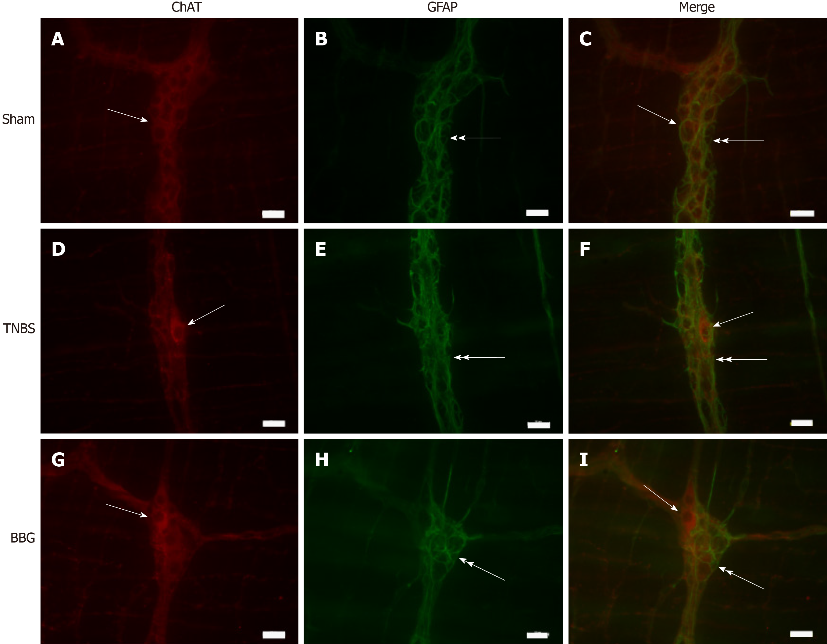
Figure 8 Double labeling of choline acetyltransferase with glial fibrillary acidic protein in the rat ileum myenteric plexus in the sham, 2,4,6-trinitrobenzene sulfonic acid and brilliant blue G groups.
A-C: Sham group; D-F: 2,4,6-trinitrobenzene group; G-I: Brilliant blue G group. Choline acetyltransferase immunoreactivity (red; A, D, and G) did not colocalize with glial fibrillary acidic protein immunoreactivity (green; B, E and H). Single arrows indicate choline acetyltransferase-positive neurons, and double arrows indicate enteric glial cell positivity. Scale bars = 50 µm. ChAT: Choline acetyltransferase; GFAP: Glial fibrillary acidic protein; TNBS: 2,4,6-trinitrobenzene sulfonic acid; BBG: Brilliant blue G.
- Citation: Souza RF, Evangelinellis MM, Mendes CE, Righetti M, Lourenço MCS, Castelucci P. P2X7 receptor antagonist recovers ileum myenteric neurons after experimental ulcerative colitis. World J Gastrointest Pathophysiol 2020; 11(4): 84-103
- URL: https://www.wjgnet.com/2150-5330/full/v11/i4/84.htm
- DOI: https://dx.doi.org/10.4291/wjgp.v11.i4.84


