Copyright
©2010 Baishideng Publishing Group Co.
World J Gastrointest Pathophysiol. Dec 15, 2010; 1(5): 154-165
Published online Dec 15, 2010. doi: 10.4291/wjgp.v1.i5.154
Published online Dec 15, 2010. doi: 10.4291/wjgp.v1.i5.154
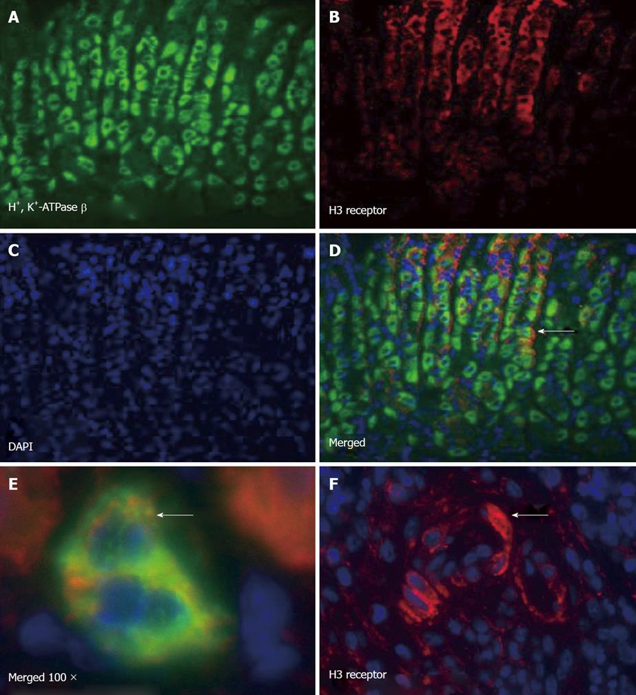
Figure 6 H3 receptor expression of mouse parietal and immune cells.
Immunofluorescence of paraffin sections of mouse stomach stained with (A) H+, K+-ATPase β subunit (FITC), (B) H3 receptor (Texas red), (C) DAPI, and (D) merged image showing colocalization; E: High power image showing parietal cells expressing the H3 receptor; F: H3 receptor expressing cells within the inflammatory infiltrate in H. felis infected mice.
-
Citation: Zavros Y, Mesiwala N, Waghray M, Todisco A, Shulkes A, Merchant JL. Histamine 3 receptor activation mediates inhibition of acid secretion during
Helicobacter -induced gastritis. World J Gastrointest Pathophysiol 2010; 1(5): 154-165 - URL: https://www.wjgnet.com/2150-5330/full/v1/i5/154.htm
- DOI: https://dx.doi.org/10.4291/wjgp.v1.i5.154


