Copyright
©2010 Baishideng Publishing Group Co.
World J Gastrointest Pathophysiol. Dec 15, 2010; 1(5): 154-165
Published online Dec 15, 2010. doi: 10.4291/wjgp.v1.i5.154
Published online Dec 15, 2010. doi: 10.4291/wjgp.v1.i5.154
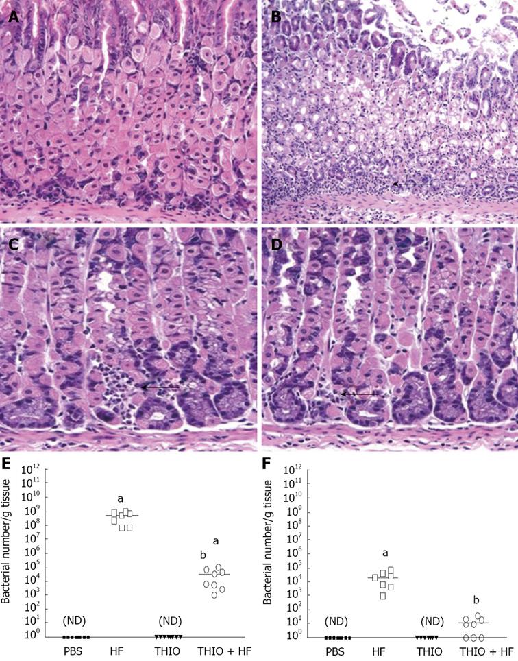
Figure 2 Thioperamide prevention of Helicobacter felis gastritis is mediated by blockade of the H3 receptor.
H&E stains of (A) PBS, (B) H. felis (HF), (C) thioperamide (THIO) and (D) THIO plus HF treated mice (magnification 400 ×). Quantification by qRT-PCR of the amount of H. felis colonizing PBS, HF, THIO and THIO plus HF treated mice. Quantitative RT-PCR was performed on RNA isolated from (E) fundic and (F) antral gastric tissue. The log fluorescent emission measured continuously during PCR per cycle was determined. Early amplification showed increased ‘starting’ fluorescent emission and was used to compute the number of Helicobacter/g tissue for each mouse using a standard curve generated from known quantities of H. felis. Scatter plots showing the bacterial count for each mouse is shown. The mean indicated by a horizontal bar for n = 8 mice. aP < 0.05 vs PBS mice; bP < 0.05 vs H. felis mice (unpaired -test); ND: non-detectable.
-
Citation: Zavros Y, Mesiwala N, Waghray M, Todisco A, Shulkes A, Merchant JL. Histamine 3 receptor activation mediates inhibition of acid secretion during
Helicobacter -induced gastritis. World J Gastrointest Pathophysiol 2010; 1(5): 154-165 - URL: https://www.wjgnet.com/2150-5330/full/v1/i5/154.htm
- DOI: https://dx.doi.org/10.4291/wjgp.v1.i5.154


