
















©2010 Baishideng.
World J Gastrointest Pathophysiol. Aug 15, 2010; 1(3): 106-108
Published online Aug 15, 2010. doi: 10.4291/wjgp.v1.i3.106
Published online Aug 15, 2010. doi: 10.4291/wjgp.v1.i3.106
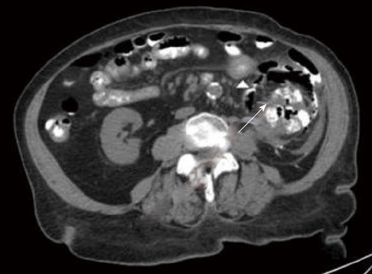
Figure 1 This image illustrates the reflux of the oral contrast into the left kidney via the colo-renal fistula, as well as free air in the renal parenchyma and perinephric space (arrow).
Also noted is thickened small bowel wall adjacent to the left kidney (arrow tip).
- Citation: Wysocki JD, Joshi V, Eiser JW, Gil N. Colo-renal fistula: An unusual cause of hematochezia. World J Gastrointest Pathophysiol 2010; 1(3): 106-108
- URL: https://www.wjgnet.com/2150-5330/full/v1/i3/106.htm
- DOI: https://dx.doi.org/10.4291/wjgp.v1.i3.106