Copyright
©2013 Baishideng Publishing Group Co.
World J Gastrointest Pathophysiol. Aug 15, 2013; 4(3): 59-62
Published online Aug 15, 2013. doi: 10.4291/wjgp.v4.i3.59
Published online Aug 15, 2013. doi: 10.4291/wjgp.v4.i3.59
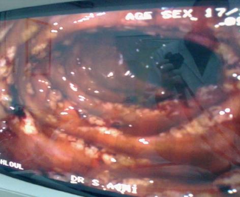
Figure 1 Colonoscopy showed multiple millimetric nodular hyperemic lesions on the mucosa.
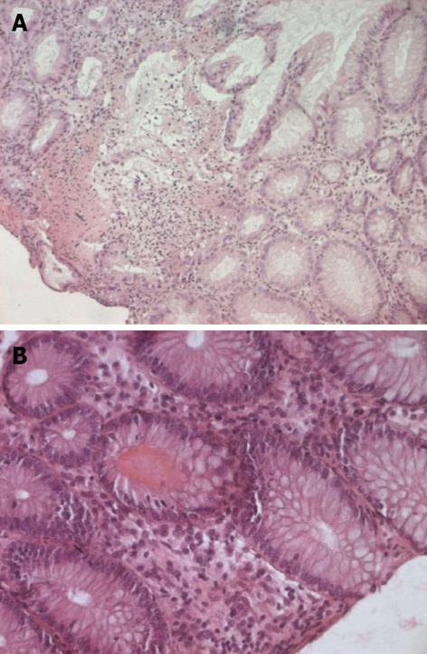
Figure 2 Mucosal biopsy of colon.
A: A well-preserved crypt structure with lymphocytes infiltration in the lamina propria (original magnification × 100); B: Viral inclusions and apoptotic bodies were absent (original magnification × 200).
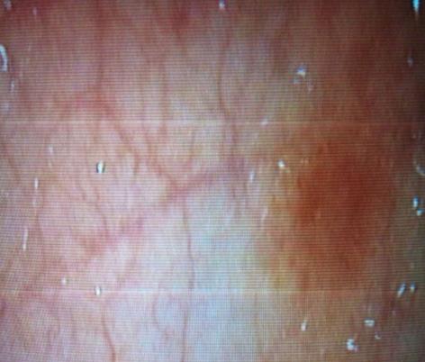
Figure 3 Macroscopic findings include erythema, edema and friability.
- Citation: Kmira Z, Nesrine BS, Houneida Z, Wafa BF, Aida S, Yosra BY, Monia Z, Sriha B, Abderrahim K. Severe hemorrhagic colitis in a patient with chronic myeloid leukemia in the blastic phase after dasatinib use. World J Gastrointest Pathophysiol 2013; 4(3): 59-62
- URL: https://www.wjgnet.com/2150-5330/full/v4/i3/59.htm
- DOI: https://dx.doi.org/10.4291/wjgp.v4.i3.59


