Copyright
©The Author(s) 2017.
World J Radiol. Jun 28, 2017; 9(6): 253-268
Published online Jun 28, 2017. doi: 10.4329/wjr.v9.i6.253
Published online Jun 28, 2017. doi: 10.4329/wjr.v9.i6.253
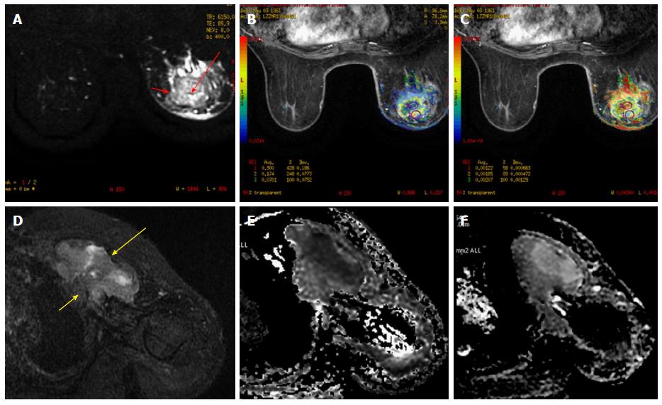
Figure 3 A 51-year-old female presented with a lump in the left breast.
A: FS T2 weighted images revealed a well-defined rounded mass with smooth margins in the left breast with central hyperintensity (long red arrow) and a hypointense rim (short red arrow). Diffusion weighted imaging (B) revealed restricted diffusion in the periphery of the lesion (ROI 1) with reduced ADC values (C). The center of the lesion showed facilitated diffusion suggestive of necrotic areas (ROI 2). Biopsy revealed invasive ductal carcinoma; D: FS T2 weighted images revealed a well-defined heterogeneously hyperintense irregular mass in the left breast (long yellow arrow) with chest wall invasion (short yellow arrow). Diffusion weighted imaging (E) revealed restricted diffusion with reduced ADC values (F). Biopsy revealed invasive ductal carcinoma. ADC: Apparent diffusion coefficient.
- Citation: Mahajan A, Deshpande SS, Thakur MH. Diffusion magnetic resonance imaging: A molecular imaging tool caught between hope, hype and the real world of “personalized oncology”. World J Radiol 2017; 9(6): 253-268
- URL: https://www.wjgnet.com/1949-8470/full/v9/i6/253.htm
- DOI: https://dx.doi.org/10.4329/wjr.v9.i6.253


