Copyright
©The Author(s) 2017.
World J Radiol. May 28, 2017; 9(5): 230-244
Published online May 28, 2017. doi: 10.4329/wjr.v9.i5.230
Published online May 28, 2017. doi: 10.4329/wjr.v9.i5.230
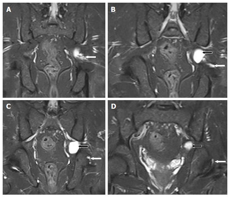
Figure 10 Images A-D show serial, T2-weighted fat suppressed coronal sections of the pelvis, demonstrate a cyst (black arrows) at the level of the left sciatic notch.
The cyst extends along the articular branch of the sciatic nerve (white arrows) and communicates with the posteromedial aspect of the ipsilateral hip joint (D). No obvious labral or capsular tear or degeneration of joint is noted.
- Citation: Panwar J, Mathew A, Thomas BP. Cystic lesions of peripheral nerves: Are we missing the diagnosis of the intraneural ganglion cyst? World J Radiol 2017; 9(5): 230-244
- URL: https://www.wjgnet.com/1949-8470/full/v9/i5/230.htm
- DOI: https://dx.doi.org/10.4329/wjr.v9.i5.230


