Copyright
©The Author(s) 2017.
World J Radiol. May 28, 2017; 9(5): 230-244
Published online May 28, 2017. doi: 10.4329/wjr.v9.i5.230
Published online May 28, 2017. doi: 10.4329/wjr.v9.i5.230
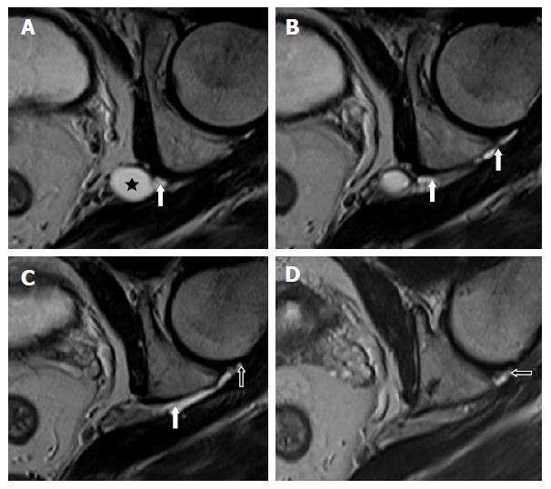
Figure 9 Image A-D show serial, T2-weighted, fast spin echo axial sections of the left hip joint highlighting a cyst (star) at the level of the left sciatic notch.
An extension along the expected course of the articular branch of the sciatic nerve (white arrows) communicating with the posteromedial aspect of the ipsilateral hip joint (open arrows) is also seen.
- Citation: Panwar J, Mathew A, Thomas BP. Cystic lesions of peripheral nerves: Are we missing the diagnosis of the intraneural ganglion cyst? World J Radiol 2017; 9(5): 230-244
- URL: https://www.wjgnet.com/1949-8470/full/v9/i5/230.htm
- DOI: https://dx.doi.org/10.4329/wjr.v9.i5.230


