Copyright
©The Author(s) 2017.
World J Radiol. May 28, 2017; 9(5): 230-244
Published online May 28, 2017. doi: 10.4329/wjr.v9.i5.230
Published online May 28, 2017. doi: 10.4329/wjr.v9.i5.230
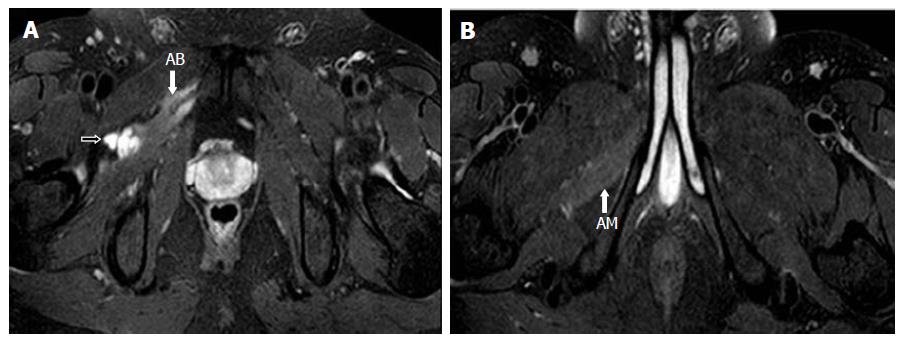
Figure 8 Image A, B show serial, T2-weighted fat suppressed axial images of the pelvis that demonstrates the further inferior extension of the cyst along the anterior branch of the obturator nerve (black arrow).
Note the denervation atrophy of adductor brevis (AB) and magnus (AM) muscles. Reprinted with permission from Acta Neurologica Belgica.
- Citation: Panwar J, Mathew A, Thomas BP. Cystic lesions of peripheral nerves: Are we missing the diagnosis of the intraneural ganglion cyst? World J Radiol 2017; 9(5): 230-244
- URL: https://www.wjgnet.com/1949-8470/full/v9/i5/230.htm
- DOI: https://dx.doi.org/10.4329/wjr.v9.i5.230


