Copyright
©The Author(s) 2017.
World J Radiol. May 28, 2017; 9(5): 223-229
Published online May 28, 2017. doi: 10.4329/wjr.v9.i5.223
Published online May 28, 2017. doi: 10.4329/wjr.v9.i5.223
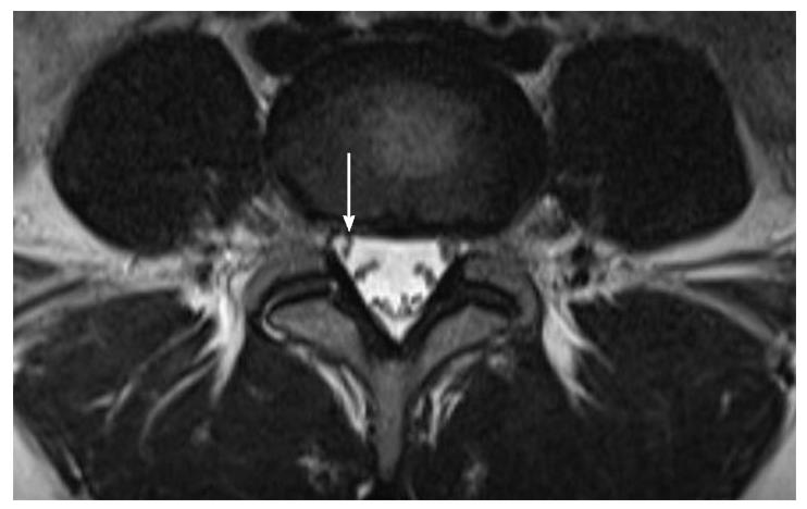
Figure 1 Axial T2-weighted magnetic resonance image of lumbar level L4/5 shows the lateral recess that is bordered laterally by the pedicle, posteriorly by the superior articular facet, and anteriorly by the vertebral body, endplate margin, and disc margin.
- Citation: Splettstößer A, Khan MF, Zimmermann B, Vogl TJ, Ackermann H, Middendorp M, Maataoui A. Correlation of lumbar lateral recess stenosis in magnetic resonance imaging and clinical symptoms. World J Radiol 2017; 9(5): 223-229
- URL: https://www.wjgnet.com/1949-8470/full/v9/i5/223.htm
- DOI: https://dx.doi.org/10.4329/wjr.v9.i5.223


