
















©The Author(s) 2017.
World J Radiol. Apr 28, 2017; 9(4): 178-190
Published online Apr 28, 2017. doi: 10.4329/wjr.v9.i4.178
Published online Apr 28, 2017. doi: 10.4329/wjr.v9.i4.178
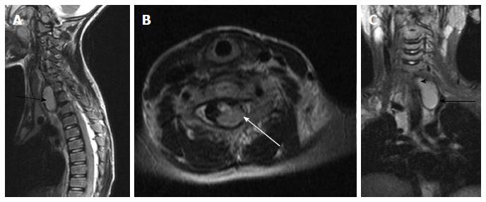
Figure 20 Neuroenteric cyst.
T2 weighted images of cervicodorsal spine in sagittal (A), axial (B) and coronal (C) planes show a bilobed lesion with both extraspinal (black arrow) and intraspinal extramedullary component (white arrow). The communication between them (arrowhead) is better appreciated in the coronal plane (C).
- Citation: Kumar J, Afsal M, Garg A. Imaging spectrum of spinal dysraphism on magnetic resonance: A pictorial review. World J Radiol 2017; 9(4): 178-190
- URL: https://www.wjgnet.com/1949-8470/full/v9/i4/178.htm
- DOI: https://dx.doi.org/10.4329/wjr.v9.i4.178