Copyright
©The Author(s) 2017.
World J Radiol. Apr 28, 2017; 9(4): 155-177
Published online Apr 28, 2017. doi: 10.4329/wjr.v9.i4.155
Published online Apr 28, 2017. doi: 10.4329/wjr.v9.i4.155
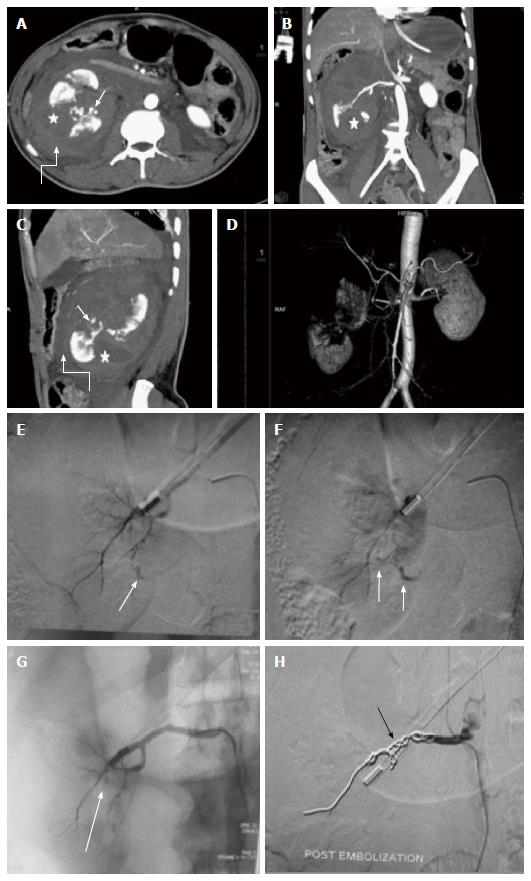
Figure 11 Main renal artery injury.
A-D: CECT in a 22 years old male following road traffic accident revealed multiple deep lacerations (asterisk) involving entire thickness of right kidney resulting in shattered kidney. Few areas of active contrast extravasation (thin arrow) were noted at the edge of laceration alongwith presence of extensive pernephric hematoma (curved arrow). Although, main renal artery was showing good luminal opacification, however, less than 25 % remaining functioning renal parenchyma precluded salvagability. Due to high post operative morbidity, angiography was resorted over nephrectomy; E, F: Selective catheterisation of main renal artery showed multiple areas of active contrast extravasation (arrow); G: Subsequently, in view of potentially life threatening haemorrhage and non-salvagable kidney, coil embolization (arrow) of main renal artery was contemplated; H: Post embolization angiography showed opacification of only proximal stump of renal artery. Following conservative management, patient was then discharged 8 d later. CECT: Contrast enhanced computed tomography.
- Citation: Singh A, Kumar A, Kumar P, Kumar S, Gamanagatti S. “Beyond saving lives”: Current perspectives of interventional radiology in trauma. World J Radiol 2017; 9(4): 155-177
- URL: https://www.wjgnet.com/1949-8470/full/v9/i4/155.htm
- DOI: https://dx.doi.org/10.4329/wjr.v9.i4.155


