Copyright
©The Author(s) 2017.
World J Radiol. Apr 28, 2017; 9(4): 155-177
Published online Apr 28, 2017. doi: 10.4329/wjr.v9.i4.155
Published online Apr 28, 2017. doi: 10.4329/wjr.v9.i4.155
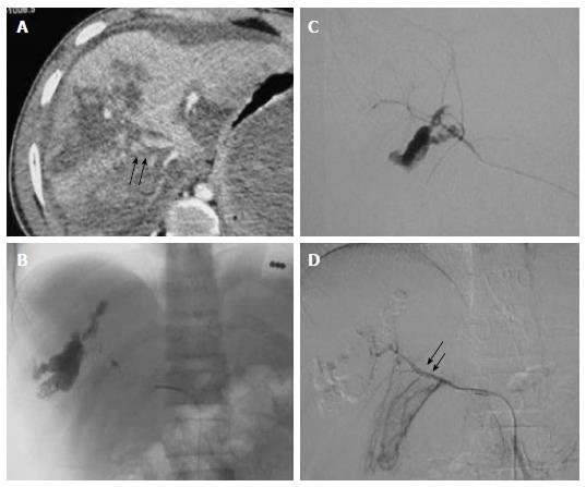
Figure 2 High grade liver injury with active contrast extravasation.
Case of blunt trauma abdomen, FAST positive, hemodynamically stable (A) CECT abdomen showed extensive laceration, intraparenchymal hematoma consistent with Grade V liver injury alongwith diffuse active contrast extravasation (arrow) within the lacerated segment VIII (arrow) of liver with resultant hemoperitoneum (B) Selective hepatic artery angiogram revealed active contrast extravasation from anterior division of right hepatic artery with increased conspicuity in (C) superselective right hepatic artery angiogram which was subsequently embolized by microcoils (D) Post embolization angiogram revealed faint opacification of branches of anterior division of RHA (arrow) with subsidence of extravasation. FAST: Focused assessment with sonography in trauma; RHA: Right hepatic artery; CECT: Contrast enhanced computed tomography.
- Citation: Singh A, Kumar A, Kumar P, Kumar S, Gamanagatti S. “Beyond saving lives”: Current perspectives of interventional radiology in trauma. World J Radiol 2017; 9(4): 155-177
- URL: https://www.wjgnet.com/1949-8470/full/v9/i4/155.htm
- DOI: https://dx.doi.org/10.4329/wjr.v9.i4.155


