Copyright
©The Author(s) 2017.
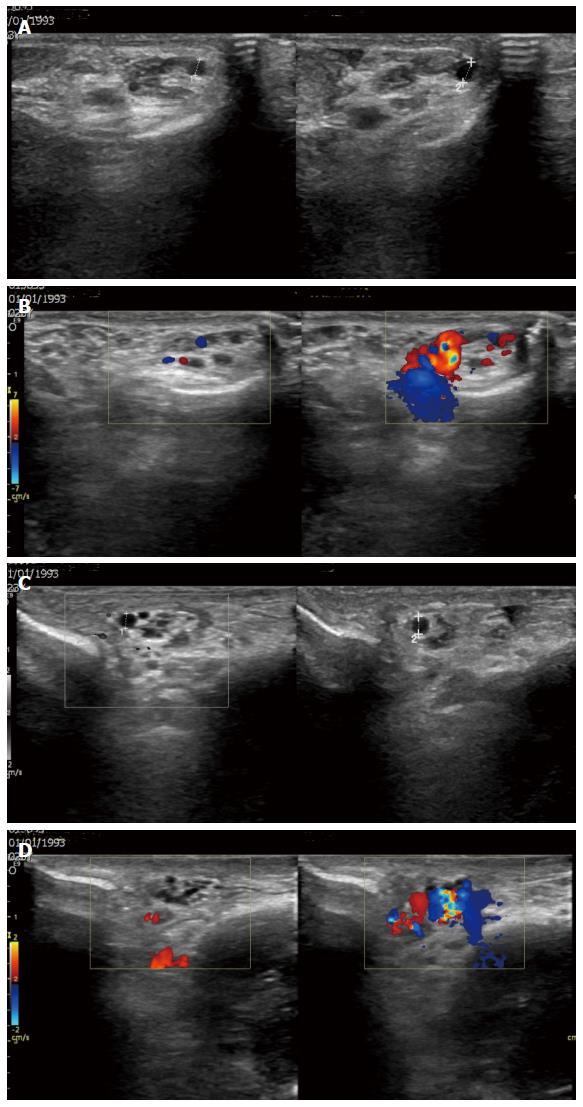
Figure 1 A 24-year-old man with bilateral varicocele.
A: Gray-scale sonographic images, longitudinal sections at the supratesticular region of the left hemiscrotum at rest and during the Valsalva maneuver. The maximal diameter of the left spermatic veins is 2.5 mm at rest and 3.5 mm during the Valsalva maneuver; B: Color Doppler sonographic images, longitudinal sections same level show blood flow reversal after Valsalva maneuver; C: Gray-scale sonographic images, longitudinal sections at the right supratesticular region. The maximal diameter of the right spermatic veins is 2.3 mm at rest and 2.8 mm during the Valsalva maneuver; D: Color Doppler sonographic images, longitudinal sections show flow reversal with Valsalva maneuver.
- Citation: Tsili AC, Xiropotamou ON, Sylakos A, Maliakas V, Sofikitis N, Argyropoulou MI. Potential role of imaging in assessing harmful effects on spermatogenesis in adult testes with varicocele. World J Radiol 2017; 9(2): 34-45
- URL: https://www.wjgnet.com/1949-8470/full/v9/i2/34.htm
- DOI: https://dx.doi.org/10.4329/wjr.v9.i2.34


